
Page 59 - Read Online
P. 59
of the “downslope” loads created by the anatomy of the Check reins
articular surface of the distal radius. Treatment of proximal interphalangeal (PIP) joint
contractures is often reported to be less than optimal.
[8]
Stenosing tenosynovitis or trigger finger The volar plate at the PIP joint is a unique structure that
The effects of the interaction between synovium and prevents hyperextension at the PIP joint and absorbs
collagen can be seen in trigger fingers. In stenosing enormous compression loads. The PIP volar plate is
tenosynovitis of the finger flexors, there is a thickened a thick, strong fibrocartilaginous structure, lined on
retinaculum or pulley that constricts the osseofibrous the volar surface by peritendinous synovium of the
[1]
tunnel through which the tendon runs. Chronic fibro‑osseous sheath and on the dorsal surface by joint
synovial irritation affects collagen deposition in the synovium. These 2 layers of synovium lie on either side
A1 pulley and leads to a progressive thickening and of the thin joint capsule at the lateral sides of the volar
sometimes metaplasia of the pulley [1‑3] [Figure 1]. During plate. Inflammation of these two different synovial
[9]
sleep, edema collects in the tendon proximal and surfaces influence each other and produce the unusual
distal to the pulley. The symptomatic sequelae include abnormal collagen hypertrophy termed the checkrein
stiffness in the mornings as patients open and close ligaments. These do not exist in the normal state but are
their fingers to “milk” the fluid back into the natural produced under the influence of this “synovial sandwich.”
shape of the tendons, or “locking” of the fingers When treating contractures of the PIP joints, one must
if a nodule is too big to pass through the pulley. release these pathological structures in order to increase
Conservative treatment may include steroid injections, the movement in the joint. Results of a study using this
splinting and activity modification. If this fail, surgery technique indicated full intraoperative extension in 110
[4]
is indicated. A release of the A1 pulley increases of 115 joints, with 2 joints requiring a collateral ligament
space to allow normal tendon gliding. Surgery has release. Three of the 115 digits required a second
been shown to be more successful in the absence of checkrein release after intraoperative gains were not
diabetes. [5] maintained. [9]
The collasyn theory explains why there is an increased Peripheral arthritis
incidence of stenosing tenosynovitis (trigger finger) in the Peripheral arthritis is secondary to synovial traction
thumb and little finger following carpal tunnel surgery. and inflammation. Osteophytes and abnormal cartilage
Infection can move from thumb to little finger through build up on the joint periphery. It is hypothesized that
the common synovial lining between the thumb, the the areas of synovial attachment are responsible for the
carpal tunnel and the fibro‑osseous sheath of the little synovitic influence on collagen and bone formation.
finger flexors. Surgery on the carpal tunnel produces With chronic synovial inflammation, the mechanical
inflammation of this communicating synovium, which traction at the synovial attachment point may play a
then has its hypertrophic effect on the collagen of the A1 part but the inflamed synovium communicates with
pulleys. the bone collagen resulting in osteophyte formation.
Fourth extensor compartment synovitis Resecting these bone areas along with excision of
Collasyn pathology is also seen in the extensor involved synovium results in clearing of the patient’s
retinaculum. Patients with fourth extensor compartment symptoms and significantly extending joint longevity.
stenosing tenosynovitis develop a thickened retinaculum. This occurs without having altered joint mechanics at
In performing ultrasound evaluation, Zhou et al. found the time of surgery.
[6]
that with increased extension of the wrist, the contact Distal radioulnar joint
area between the extensor retinaculum and the extensor This approach has been used in the treatment of
tendons decreased, causing increased friction. We have arthritis in the distal radioulnar joint (DRUJ). The
found that a release of the septa between the fourth treatment of DRUJ degenerative arthritis following
and fifth extensor compartments without releasing the failure of conservative treatment such as splinting
external retinaculum is all that is needed to provide and antiinflammatory medication includes complete
sufficient room for the tendons. [7] elimination of the arthritic joint, as popularized by
[10]
[11]
Darrach, a hemiresection‑interposition technique, the
[12]
matched distal ulna resection or the Sauvé‑Kapandji
procedure, as well as ulnar head or total joint
[13]
replacement. A modified DRUJ arthritis technique
based on the concept of proximal to distal progression
of degenerative joint disease at the DRUJ has been
[14]
described [Figure 2]. The proximal one‑third to
one‑half of the articular surface is typically resected
around the entire circumference of the joint. In
one published study, all patients noted significant
[14]
improvement in symptoms. One patient went on to
have a matched ulna arthroplasty. In another report on
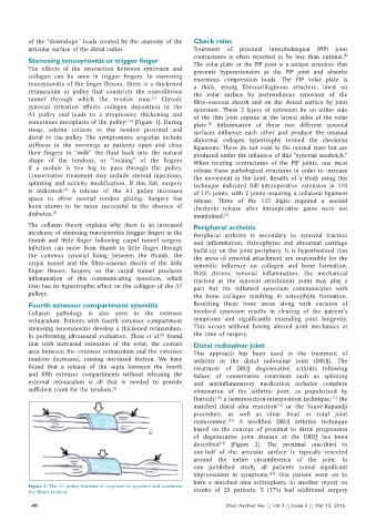
Figure 1: The A1 pulley thickens in response to synovitis and constricts
the flexor tendons results of 29 patients, 5 (17%) had additional surgery
48 Plast Aesthet Res || Vol 2 || Issue 2 || Mar 13, 2015