
Page 55 - Read Online
P. 55
a b c d
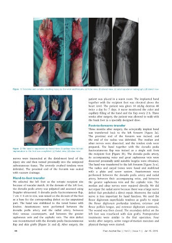
Figure 1: Extensive and complex crushing injury to the wrist and forearm. (a) Volar view; (b) dorsal view; (c) anterioposterior radiograph; (d) lateral view
patient was placed in a warm room. The implanted hand
together with the recipient foot was elevated above the
heart level. The patient was given 10 mL/kg dextran 40
twice a day for 7 days. A nurse monitored the color and
capillary filling of the hand and the flap every 2 h. Three
weeks after surgery, the patient was allowed to walk with
a b the bank foot in a specially designed shoe.
Foot‑to‑forearm transfer
Three months after surgery, the ectopically implant hand
was transferred back to the left forearm [Figure 3a].
The proximal end of the forearm was incised, and
the end of the radius was debrided. The median and
ulnar nerves were dissected, and the tendon ends were
c d
Figure 2: The hand is amputated. (a) Dorsal view; (b) palmar view. Ectopic prepared. The hand together with the dorsalis pedis
implantation to the foot was completed. (c) Radial view; (d) ulnar view fasciocutaneous flap was incised as a single unit from
the recipient foot [Figure 3b]. The dorsalis pedis artery,
nerves were transected at the distal‑most level of the its accompanying veins and great saphenous vein were
injury site and then turned proximally into the uninjured dissected proximally until suitable lengths were obtained.
subcutaneous tissue. The severely crushed tendons were The hand was transferred to the left forearm [Figure 3c‑e].
debrided. The proximal end of the forearm was sealed The radius and carpal bones were fused and stabilized
with vacuum drainage. with a plate and screw system. Anastomoses were
performed between the dorsalis pedis artery and radial
Hand‑to‑foot transfer artery, between their accompanying veins, and between
We selected the left foot as the ectopic recipient site the greater saphenous vein and the cephalic vein. The
because of vascular match. At the dorsum of the left foot, median and ulnar nerves were repaired directly. We did
the dorsalis pedis artery was palpated and assessed using not repair the radial nerve because there was a large nerve
Doppler ultrasound. A dorsalis pedis fasciocutaneous flap, defect that precluded a direct repair. Moreover, the radial
7 cm × 8 cm in size, was raised on the dorsum of the foot nerve is less important for hand function. We used the
as a base for the corresponding defect on the amputated flexor digitorum superficialis tendons as grafts to repair
part. The hand was stabilized to the tarsal bones with the flexor digitorum profundus tendons, extensor and
K‑wires. Anastomoses were performed between the flexor pollicis longus, and extensor digitorum communis.
dorsalis pedis artery and the radial artery, between The wound was then closed. The secondary defect on the
their venous counterparts, and between the greater left foot was resurfaced with skin grafts. Postoperative
saphenous vein and the cephalic vein. The skin defect treatments were similar to the first operation. Four
was reconstructed with the dorsalis pedis fasciocutaneous weeks after surgery, active range‑of‑motion exercises and
flap and skin grafts [Figure 2c and d]. After surgery, the physical therapy were started.
44 Plast Aesthet Res || Vol 2 || Issue 1 || Jan 15, 2015