Page 50 - Read Online
P. 50

and antihelix, a wedge or star‑shaped excision technique is
                                                              a preferable option.  It consists of a full‑thickness excision
                                                                               [5]
                                                              of skin and cartilage with the apex pointing to the anterior
                                                              surface of the ear and extending to the conchal area. When
                                                              designing the wedge, it is important to define an apex
                                                              angle smaller than 30°.  The resulting wound is then closed
                                                                                 [6]
                                                              primarily in layers, with the cartilage secured by long lasting
                                                              sutures.  It is helpful, when possible, to use an offset skin
                                                                     [5]
                                                              closure around the rim. To decrease the risk of rim notching,
                                                              the skin should not be approximated and secured over the
                                                              cartilage space.  Usually, the ear is shortened slightly while
                                                                           [7]
                                                              maintaining the premorbid contour.  The advantages of
                                                                                             [7]
                                                              wedge resection are: a one‑stage operation, simple and fast
                                                              dissection, and minimal resultant scar.
                                                              However, the limitation  of this technique is that it can
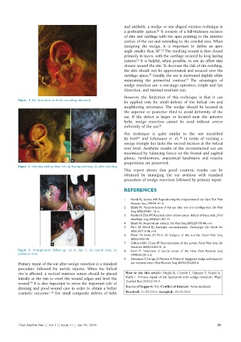
          Figure 1: Ear laceration on helix extending anteriorly
                                                              be  applied only for small defects of the  helical rim  and
                                                              neighboring  structures.  The  wedge  should be  located in
                                                              the  superior or posterior third to avoid deformity  of the
                                                              ear. If the defect is larger or located near the anterior
                                                              helix,  wedge resection cannot be used without severe
                                                              deformity of the ear. [2]
                                                              Our  technique  is  quite  similar to  the  one  described
                                                                                          [9]
                                                                     [8]
                                                              by  Ferri  and Schonauer  et  al.,  in terms  of excising  a
                                                              wedge triangle but lacks the second incision at the helical
                                                              root  level.  Aesthetic  results  of  the  reconstructed ear  are
                                                              maximized by balancing forces on the frontal and sagittal
                                                              planes. Furthermore, anatomical landmarks  and relative
           a                        b
                                                              proportions are preserved.
          Figure 2: Stitching with prolene 5‑0. (a) During stitching, (b) after stitching
                                                              This report shows that good cosmetic results can be
                                                              obtained by managing  the ear avulsion with standard
                                                              procedure of wedge resection followed by primary repair.


                                                              REFERENCES

                                                              1.   Havlik RJ, Sadove AM. Repositioning the malpositioned ear. Oper Tech Plast
                                                                  Reconstr Surg 1997;4:141‑5.
                     a             b                          2.   Elsahy NI. Reconstruction of the ear after skin and cartilage loss. Clin Plast
                                                                  Surg 2002;29:201‑12, vi.
                                                              3.   Reddy LV, Zide MF. Reconstruction of skin cancer defects of the auricle. J Oral
                                                                  Maxillofac Surg 2004;62:1457‑71.
                                                              4.   Elsahy NI. Acquired ear defects. Clin Plast Surg 2002;29:175‑86, v‑vi.
                                                              5.   Park  SS,  Hood  RJ. Auricular  reconstruction.  Otolaryngol  Clin  North Am
                                                                  2001;34:713‑38, v‑vi.
                                                              6.   Pham TV,  Early  SV,  Park  SS.  Surgery  of  the  auricle.  Facial Plast Surg
                                                                  2003;19:53‑74.
                           c                                  7.   Calhoun KH, Chase SP. Reconstruction of the auricle. Facial Plast Surg Clin
                                                                  North Am 2005;13:231‑41, vi.
          Figure  3:  Postoperative  follow‑up.  (a) At  day 7,  (b) lateral view,  (c)   8.   Ferri  M. Treatment of partial losses of the helix.  Plast Reconstr Surg
          posterior view                                          1998;101:2011‑2.
                                                              9.   Schonauer F, Campa D, Monaco A, Molea G. Staggered wedge technique for
          Primary repair of the ear after wedge resection is a standard   ear reconstruction. Plast Reconstr Surg 2010;125:e203‑4.
          procedure followed for auricle injuries. When the helical
          rim is affected, a vertical mattress suture should be placed   How to cite this article: Singla B, Chawla I, Gautam P, Goyal A,
          initially at the rim to evert the wound edges and level the   Rathi J. Primary repair of ear laceration with wedge resection. Plast
                                                               Aesthet Res 2015;2:38-9.
          wound.  It is also important to stress the important role of
                [4]
          dressing and good wound care in order to obtain a better   Source of Support: Nil, Conflict of Interest: None declared.
          cosmetic outcome. [1,5]  For small composite defects of helix   Received: 21-05-2014; Accepted: 10-10-2014


          Plast Aesthet Res || Vol 2 || Issue 1 ||  Jan 15, 2015                                            39
   45   46   47   48   49   50   51   52   53   54   55