Page 353 - Read Online
P. 353
Table 1: Modified Lane and Sandhu radiological scores, mean new bone formation in Histology (%), and mean
          compressive strength (MPa) of the rabbit’s radius in each group at 3 months after surgery
           Group        Scaffold implantation       Mean radiological      Mean compressive       Mean new bone
                                                         scores             strength (MPa)        in Histology (%)
           1            HDCB only                      2.95 ± 0.58            31.14 ± 6.72          29.60 ± 8.33
           2            Periosteum-wrapped HDCB        5.57 ± 0.51            73.00 ± 7.20          49.79 ± 11.69
           3            HDCB/BM-MSCs                   6.41 ± 1.03            80.57 ± 8.50          64.12 ± 11.31
           4            Periosteum-wrapped HDCB/       8.58 ± 0.64            129.31 ± 5.99         80.50 ± 4.96
                        BM-MSCs
           HDCB: Human demineralized cancellous bone, BM‑MSCs: Bone marrow mesenchymal stem cells
                                                              source, which is more convenient for isolation and
                                                              expansion  when  compared with  periosteum‑derived  cells.
                                                              To  further  enhance  the  current  bone  tissue  engineering
                                                              strategies, a successful cellular replacement for  periosteum
                                                              or tissue‑engineered periosteum should be investigated.
                                                              Zhang et al.  previously reported successful regeneration
                                                                        [11]
                                                              of segmental  bone defects in rabbit  ulnas using
                                                              periosteum encapsulated  scaffolds seeded with MSCs,
                                                              with  an  increase  in  the  newly  formed bone  area  to
                                                              80.1% ± 9.6%. This result is compatible with the results of
                                                              the current study at 80.5% ± 4.96%.

                                                              Xenogeneic demineralized cancellous bone grafts, which
                                                              have  the  advantages  of  favorable  cellular  compatibility
                                                              and histocompatibility as a scaffold, have widely been
                                                              used for the repair of short bony defects showing the
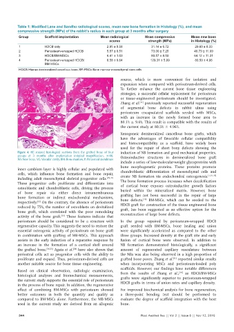
          Figure  4:  HE  stained  histological  sections  from  the  grafted  bone  of  four   induction of NB formation and good mechanical properties.
          groups  at  3  months  after  implantation  (original  magnification,  ×40).   Osteoinductive structures in demineralized bone graft
          NB: New bone, VC: Vascular cavity, BM: Bone marrow, P: Periosteal membrane
                                                              include a series of low‑molecular‑weight glycoproteins with
                                                              bone morphogenetic proteins. These proteins promote
          inner cambium layer is highly cellular and populated with   chondroblastic differentiation of mesenchymal cells and
          cells, which influence bone formation and  bone  repair,   create NB formation via endochondral osteogenesis. [1,31,35]
          including adult mesenchymal skeletal progenitor cells. [29,31]    The bone formation process increases when decalcification
          These progenitor cells proliferate and differentiate into   of  cortical  bone  exposes  osteoinductive  growth  factors
          osteoblastic and chondroblastic cells, driving the process   buried within the mineralized matrix. However, bone
          of bone repair via either direct intramembranous    grafting  has not  been  successful in  the  repair of  large
          bone formation or indirect endochondral mechanisms,   bone  defects.   BM‑MSCs,  which  can  be  seeded  to  the
                                                                          [13]
                    [32]
          respectively.  On the contrary, the absence of periosteum   HDCB graft for construction of the tissue engineered bone
          reduced by 75%, the number of osteoblasts on devitalized   graft, has been suggested as an effective option for the
          bone graft, which correlated with the poor remodeling   reconstruction of large bone defects.
          activity of the bone graft.  These features indicate that
                                [33]
          periosteum should be considered to be a structure with   In  the  group repaired  by  periosteum‑wrapped  HDCB
          regenerative capacity. This suggests the need to restore the   graft seeded with  BM‑MSCs,  bone healing  and union
          essential osteogenic activity of periosteum on bone graft   were  significantly  accelerated as  compared  to  the  other
          in combination with grafting of MB‑MSCs. This approach   three groups. Increased density at the graft site and early
          assists  in  the  early  induction  of  a  reparative  response  by   fusion  of  cortical bone  were  observed.  In  addition to
          an increase in the formation of a cortical shell around   NB formation demonstrated histologically, a significant
          the grafted bone. [34,35]  Agata et al.  have also shown that   amount of regenerated capillary vasculature between
                                      [34]
          periosteal cells act as  progenitor cells with the ability to   the NBs was also being observed in a high proportion of
                                                                                         [11]
          proliferate and expand. Thus, periosteum‑derived cells are   grafted bone pores. Zhang et al.  reported similar results
          another suitable source for bone tissue engineering.  when incorporating MSCs and periosteum‑loaded poly
                                                              scaffolds.  However, our findings have notable differences
          Based on  clinical observation,  radiologic examination,   from  the  results  of  Zhang  et  al.,   as  HDCB/BM‑MSCs
                                                                                            [11]
          histological analyses  and biomechanical  measurements,   grafts  were  significantly  superior to periosteum‑wrapped
          the current study supports the essential role of periosteum   HDCB grafts in terms of union rates and capillary density.
          in the process of bone repair. In addition, the regenerative
          effect of combining  BM‑MSCs with  periosteum  showed   For improved biochemical analysis for bone regeneration,
          better outcomes in both the quantity and quality as   a three‑point bending  test  should be  performed to
          compared  to  BM‑MSCs alone.  Furthermore,  the  MB‑MSCs   evaluate  the  degree  of scaffold  integration  with  the  host
          used in the current study are derived from an allogenic   bone.

           344                                                           Plast Aesthet Res || Vol 2 || Issue 6 || Nov 12, 2015

