Page 352 - Read Online
P. 352
the HDCB grafts were completely degraded in this    significant differences were found between Groups 2 and 3
          group [Figure 2].                                   (P  <  0.05), while  both  groups were  statistically  superior
                                                              as compared to Group 1 (P < 0.05) [Table 1].
          Radiological outcomes
          At 3  months postoperatively, there was a small amount   Biomechanical testing results
          of callus formation at the defect gaps in Group  1. NB   Radii of rabbits with partial or complete union were
          formation was found  to account for over half of the   subjected to biomechanical testing. Results of the
          material at the reconstructed bone in Groups  2 and 3.   biomechanical  tests  are  summarized  in  Table  1.  Group  4
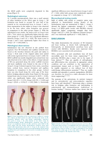
          Bone regeneration in the radius in Group 4 was observed   showed  the  highest  compressive  strength  (P  <  0.05).
          to be the best, where callus  formation was greatest   Group  3  of  HDCB  grafts  seeded  with  BM‑MSCs  showed
          in comparison to the other groups  [Figure  3]. With the   significantly  higher  compressive  strength  than  both
          radiological score results, the mean score in Group 4 was   Groups 1 and 2 (P < 0.05). The difference between Groups 1
          8.58 ± 0.64, which was significantly higher than the other   and 2 was statistically significant (P < 0.05) [Table 1].
          three groups (P < 0.05). There was a significant difference
          between Groups 2 and 3 (P < 0.05). The mean scores in   DISCUSSION
          Groups  2 and 3 were significantly  higher  than those in
          Group 1 (P < 0.05) [Table 1].                       This study demonstrates the presence of NB formation
          Histological observations                           and bone healing, as shown both radiologically  and
          Inflammation  was  not  observed in  the  grafted  bone   histologically,  on  demineralized cancellous bone  graft
          segment.  Poor NB formation  and capillary  network were   seeded with BM‑MSCs.  Results were improved when
          found  at the interface between the graft and radius in   BM‑MSCs were associated with periosteum.
          Group  1.  Both ends of the  original  radius were  united   MSCs,  periosteal cells and osteoblasts have all been
          with newly regenerated bone in Groups 2 and 3, while the   successfully used for bone tissue engineering. [4,18]  In
          HDCB scaffold was mostly degraded and cortical bone was   particular,  BM‑MSCs  play a major role in  the  repair of
          only observed at the center of the defects. A larger amount   bone  defects. [22‑25]  They  are  capable of self‑replication
          of NB was generated along the entire scaffold  structure   and  differentiation  into  osteocytes  in  appropriate  culture
          and more  capillaries  were  formed  in  the  area of NB in   conditions  and  can  contribute  to  the  regeneration  of
          Group 4. Group 4 showed superior bone union, cancellous   mesenchymal  tissues  such  as  bone. [3,26]   BM‑MSCs  can
          bone, cortical bone, marrow formation and capillary   be rapidly expanded  ex vivo without loss of their
          formation in comparison to the other groups. Cortical   multi‑lineage  differentiation  potential.  They are readily
                                                                                               [13]
          bone was also found  along the entire gap of the bone   available and amenable to genetic manipulation. BM‑MSCs
          defect, bridging adjacent native bone [Figure 4]. The newly   can, therefore, be viewed as a viable alternative for bone
          formed bone area in Group 4 increased to 80.5% ± 4.96%,   tissue engineering. [8,11,27,28]
          which was significantly higher when  compared with   The anatomy of the periosteum, its nutrient transport
          Group 3 (64.12% ± 11.31%), Group 2 (49.79% ± 11.69%) and   and its osteoinductive and  osteoconductive  capacities
          Group 1 (29.6% ± 8.33%) (P < 0.05) [Table 1]. Statistically
                                                              have been well described.  Periosteum plays a primary
                                                                                     [29]
                                                              role in bridging callus formation and participating in
                                                              endochondral  and  intramembranous  ossifications  in
                                                              fracture healing.  Previous studies have shown that the
                                                                            [30]


          a           b           c            d
          Figure 2: Gross observations of the reconstruction of radius at 3 months
          after surgery. (a) Small amount of callus and fibrous‑like tissue in the
          interspaces  between  defect  and  human  demineralized  cancellous
          bone graft in  Group  1;  (b) callus formed in  the  defect repair by   a  b  c           d
          periosteum‑wrapped human demineralized cancellous  bone graft in
          Group  2;  (c) significant amount of callus and bony union filled in the   Figure 3: Results of X‑ray at the 3 months postoperation. (a) A few calluses
          defect repair with the human demineralized cancellous  bone graft   at the defect gap in Group 1; (b) significant new bone information at the
          seeded with mesenchymal stem cells in Group  3;  (d)  complete bone   reconstructed bone  in  Group 2; (c) more  new  bone  formation  between
          healing in the defect repair by periosteum‑wrapped human demineralized   graft and bone tissue in Group 3; (d) almost remodeling of new formed
          cancellous bone graft seeded with bone marrow mesenchymal stem cells   bone along the entire gap of the bone defect in Group  4, and the
          in Group 4                                          cortical bone bridged to the adjacent native bone
          Plast Aesthet Res || Vol 2 || Issue 6 || Nov 12, 2015                                             343

