Page 351 - Read Online
P. 351
Committee  and the Ethics  Committee  of the Medical   2 (25‑50% NB formation), 3  (50‑75% NB formation) or
          Training and Research Center. The rabbit bone defect   4 (more than 75% NB formation). The score assigned to the
          model was established as described previously. [11,13]  The   assessment of union was 0 (nonunion), 1 (possible union)
          rabbit was anesthetized with a combination of intravenous   or 2 (radiographic union). The proximal and distal unions of
          sodium pentobarbital at 20 mg/kg and intramuscular ketamine   the bone graft were separately evaluated. The remodeling
          at  50  mg/kg.  The  anterolateral  side  of  the  forelimb  was   score assigned was 0  (no evidence of remodeling)
          shaved and sterilized with 10% povidone‑iodine. The radius   2  (intramedullary remodeling) or 4  (cortical remodeling).
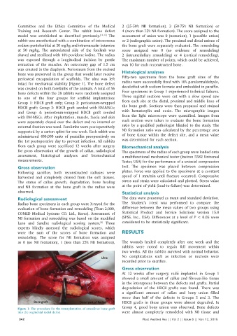
          was  exposed  through  a  longitudinal  incision  by  gentle   The maximum number of points, which could be achieved,
          retraction  of  the  muscles.  An  osteotomy  gap  of 1.5  cm   was 10 for each reconstructed bone.
          was created in the diaphysis. Periosteum from the excised
          bone was preserved in the group that would later receive   Histological analyses
          periosteal encapsulation of scaffolds.  The ulna was left   Fifty‑two  specimens  from  the  bone  graft  sites  of  the
          intact for mechanical stability [Figure 1]. The bone defect   radius were successfully fixed with 10% paraformaldehyde,
          was created on both forelimbs of the animals. A total of 56   decalcified with sodium formate and embedded in paraffin.
          bone defects within the 28 rabbits were randomly assigned   Four specimens in Group 1 experienced technical failures.
          to  one  of  the  four groups for scaffold implantation:   Three  sagittal sections were cut with a slow speed saw
          Group  1: HDCB  graft  only; Group  2: periosteum‑wrapped   from each site at the distal, proximal and middle lines of
          HDCB graft; Group  3: HDCB graft seeded with BM‑MSCs;   the bone graft. Sections were then prepared and stained
          and Group  4:  periosteum‑wrapped  HDCB graft seeded   with hematoxylin and eosin. The micrographic images
          with BM‑MSCs. After implantation, muscle, fascia and skin   from  the  light  microscope were  quantified.  Images  from
          were separately closed over the defect and no internal or   each section were taken to evaluate the bone formation
          external fixation was used. Forelimbs were postoperatively   ratio by a qualified pathologist blinded to the study. The
          supported by a carton splint for one week. Each rabbit was   NB formation ratio was calculated by the percentage area
          administered 400,000 units of penicillin preoperatively on   of bone tissue  within  the defect site,  and a mean  value
          the 1st postoperative day to prevent infection. All rabbits   was determined for each section.
          from  each group were  sacrificed  12  weeks  after  surgery   Biomechanical analysis
          for gross observation of the growth of callus, radiological   The specimens of the radius of each group were loaded onto
          assessment,  histological analyses and  biomechanical   a multifunctional mechanical tester (Instron 5582 Universal
          measurements.                                       Tester, USA) for the performance of a uniaxial compression

          Gross observation                                   test.  The specimen  was placed between  compression
          Following sacrifice,  both reconstructed radiuses were   plates. Force was applied to the specimens at a constant
          harvested and completely cleared from the soft tissues.   speed of 1  mm/min  until fracture occurred. Compressive
          The  status  of  callus growth,  degradation,  bone  healing   stress and strain were calculated and plotted. Stress value
          and  NB formation at the bone graft in the radius were   at the point of yield (load‑to‑failure) was determined.
          observed.                                           Statistical analysis

          Radiological assessment                             The data were presented as mean and standard deviation.
          Radius bone specimens in each group were X‑rayed for the   The Student’s  t‑test  was performed to compare  the
          evaluation of bone formation and remodeling (Titan 2,000,   difference between the mean values of two groups using
          COMED Medical Systems CO. Ltd., Korea). Assessment of   Statistical  Product and Service Solutions version 15.0
          NB formation and remodeling was based on the modified   (SPSS, Inc., USA). Differences at a level of P < 0.05 were
                                                   [1]
          Lane and Sandhu radiological scoring system.  Three   considered to be statistically significant.
          experts  blindly  assessed  the  radiological  scores,  which
          were the sum of the scores of bone formation and    RESULTS
          remodeling. The score for NB formation was assigned
          as 0  (no NB formation), 1  (less than 25% NB formation),   The wounds healed completely after one  week and the
                                                              rabbits were noted to regain full movement within
                                                              two weeks. All the rabbits survived with normal behavior.
                                                              No complications such as  infection  or necrosis  were
                                                              recorded prior to sacrifice.
                                                              Gross observation
                                                              At 12  weeks after surgery, radii implanted in Group  1
                                                              showed a small amount of callus and fibrous‑like tissue
                                                              in the interspaces between the defects and grafts. Partial
                                                              degradation of the HDCB grafts was found. There was
                                                              a significant amount of callus and bony union filled
                                                              more  than  half  of  the  defects  in  Groups  2  and  3.  The
                                                              HDCB grafts in these groups were almost degraded. In
          Figure 1: The procedure for the transplantation of cancellous bone graft   Group  4, good bony union  was observed. Bone defects
          into the segmental radial defect                    were almost completely remodeled with NB tissue and
           342                                                           Plast Aesthet Res || Vol 2 || Issue 6 || Nov 12, 2015

