
Page 314 - Read Online
P. 314
to suspend the platysma to the mastoid fascia. This can procedure described herein can therefore, be considered
be achieved by lifting the SCM muscle with one hand to be a repositioning of both the SMAS and fat.
and sliding a mandibular awl beneath it in an anterior The so‑called “short scar” SMAS lift, with a strictly
[19]
direction [Video 6]. The PDS suture is picked up by vertical vector, is not so short. The scar is quite long
the awl, pulled posteriorly and knotted to the mastoid because it requires an extra skin excision in the lower
fascia using a widow needle [Figure 13]. The PDS suture eyelid [Figure 15a and c] and vertical pleating in the
requires many knots to hold. The volume and ends of neck [Figure 15b] with difficult undermining in the
the suture may cause pain when they press on the skin, retroauricular area and an extra posterior hairline incision.
such as during sleep. In addition, the ends may pierce
the skin and cause painful inflammation. Suturing the The total length of these “short scar” incisions averages
surrounding tissues on the top of the knots with a 13 cm, whereas the currently proposed procedure uses an
4‑0 Vicryl suture can prevent this problem. incision with an average length of 11 cm.
The 2‑0 PDS purse string suture that is woven into the MAINTAINING TRAGUS AND EARLOBE
SMAS and picks up the remaining fat in the jowl area POSITION
is suspended to the temporoparotid fascia described
by Lore. The same procedure is used for the 2‑0 PDS Trimming of the preauricular skin should be conservative
suture loops picking up the lower side of the nasolabial because the pretragal high suture suspension creates
fold, which smooth the nasolabial groove and provide
a moderate lift to the malar prominence. However, this tension on the tragal cartilage via the SMAS. Visibility of
effect does not appear to be maintained over time. the external auditory canal is not aesthetically pleasing.
The dermis overlying the tragal cartilage is trimmed
The high suspension is achieved by suturing the dermis over 1.5 cm to recreate the pretragal groove [Figure 16].
in the pretragal area down to the parotid fascia using Otherwise, the flat appearance of the surface in front of
4‑0 Vicryl, with one stitch above and one stitch below
the level of the tragus. Another useful high suspension
maneuver was demonstrated by Dr. Heinz Bull at during
the 2006 meeting of the German Association for Aesthetic
Surgery in Düsseldorf. Using this technique, the skin
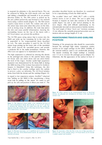
flap is fixed to the conchal cartilage under the earlobe
to prevent a pixie ear deformity. The 4‑0 Vicryl includes
tissue from both the dermis and the cartilage [Figure 14].
In regard to low suspension sutures, Hoefflin observed
[25]
that “pulling on the SMAS is like repositioning a living
room sofa by pulling on the carpet. It’s easier to just
pick up the sofa and position it where you want it”. The
Figure 12: When sutured at the temporoparotid fascia as described
by Lore, the platysma and subcutaneous fat bulge in the superolateral
esthetic zone
a b
Figure 11: The platysma is more mobile a few centimeters in front of
the sternocleidomastoid muscle than directly on top of it. (a) Prior
[21]
to elevating the platysma with the suture loop; (b) after sliding the
platysma along the superficial layer of the deep cervical fascia. Courtesy
of Dr. Daniel Labbé, Caen, France
a b a b
Figure 13: Platysma suspension. (a) A mandibular awl is used to lift the Figure 14: Suspension of the skin flap to the ear. (a) High suspension of
platysma under the sternocleidomastoid muscle; (b) suspension at the dermis to the conchal cartilage using a 4‑0 Vicryl suture; (b) the earlobe
mastoid periosteum is pushed upward and backward
Plast Aesthet Res || Vol 2 || Issue 6 || Nov 12, 2015 305