
Page 312 - Read Online
P. 312
lifting effect to the neck compared to the high suspension
of the SMAS flap used in a classic facelift. The disadvantage
is that the purse string formed by plication of the SMAS
can result in preauricular fullness in heavier patients. The
[16]
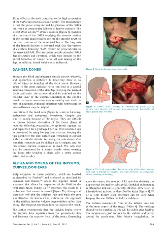
lateral SMAS ectomy offers a solution [Figure 5]. Excision
of a portion of the SMAS overlying the anterior section
of the parotid gland secures the mobile anterior SMAS to
the fixed portion of the superficial fascia. The long axis
of the lentoid incision is oriented such that the vectors
of elevation following SMAS closure lie perpendicular to
the nasolabial fold. This procedure avoids extensive SMAS
flap dissection and elevation, which risks damage to the
buccal branches of cranial nerve VII and tearing of the
flap. In addition, lateral bulkiness is addressed.
DANGER ZONES Figure 4: Open‑sky liposuction of the jowls
Because the SMAS and platysma muscle are not elevated,
and liposuction is preferred to lipectomy, there is no
risk of injury to branches of the facial nerve. However,
injury to the great auricular nerve can lead to a painful
neuroma. Dissection of the skin flap overlying the mastoid
fascia and under the earlobe should be confined to the
reticular layer of the dermis. Sensation in the earlobe
should return to normal or near normal by one week. In
case of neuralgia, repeated injections with ropivacaine or a b
betamethasone may be helpful. Figure 5: Lateral SMAS ectomy, as described by Baker (2,000).
(a) Schematic diagram; (b) intraoperative photograph. Resorbable 4‑0
Laceration of the facial vein [Figure 6] leads to bleeding, Vicryl placed after resection
ecchymosis and sometimes hematoma. Coagula can
lead to oozing because of fibrinolysis. They are difficult
to remove because dissection of the tissue planes is
required; following evacuation the epidermis appears lax
and pigmented for a prolonged period. Vein laceration can
be prevented by using Metzenbaum scissors, keeping the
tips parallel to the skin surface and remaining in contact
with the reticular dermis. Retrieving the vein stump after
complete resection can be difficult as it retracts, and for
this reason, bipolar coagulation is used. The vein may
also be punctured by a suture needle when weaving
the loops (the weaving is done with a sertix suture:
suture and needle).
ALPHA AND OMEGA OF THE INCISION,
CURVES, DOG‑EARS
Figure 6: The facial vein (darker blue) at the edge of the dissection area
(red oval) is difficult to visualize and may, therefore, be accidentally
Scalp extension to create sideburns, which are beveled nicked, resulting in bleeding
as described by Frechet and scalloped as described by
[17]
Camirand and Doucet, opens the superior tunnel in upon the vector, the amount of lift and skin elasticity, the
[18]
a manner that allows for suspension of the SMAS to the dog‑ear may be small or substantial. Cephalad undermining
temporalis fascia [Figure 7a]. However, the result is a is attempted first and is generally effective. Otherwise, an
[19]
visible scar that cannot be erased [Figure 7b]. Attempts at infra‑sideburn incision, as described by Knize [Figure 8], [5,20]
correction will thin the sideburn hair and make the area or a 1‑cm hockey stick extension can be used while
less attractive. As mentioned in section one, the approach keeping the scar hidden behind the sideburn.
to the midface involves volume augmentation rather than
lifting. The temporal extension does not improve the result. The incision descends in front of the inferior crus and
at the inner aspect of the tragus [Video 4]. The cartilage
The author recommends that the incision begins where should not be notched as this will be visible after healing.
the anterior helix separates from the preauricular skin The incision runs just anterior to the earlobe and curves
and becomes the superior helix of the pinna. Depending around its attachment. After bipolar coagulation, the
Plast Aesthet Res || Vol 2 || Issue 6 || Nov 12, 2015 303