
Page 313 - Read Online
P. 313
superior subcutaneous tunnel is prepared. The incision The 3 (sometimes 2 or 4) low suture suspension loops
then runs behind the pinna in the cephalad direction to and 3 high suspension sutures are made of the resorbable
the conchal cartilage; the scar will contract and be pulled material. The loops are woven in the SMAS layer and
down into the groove. If the incision is inadvertently provide a mild purse string action that should be taken
placed in the auriculomastoid groove, the scar will be into clinical consideration [Video 5].
pulled into the visible mastoid area. The incision becomes
horizontal at the level of the external auditory canal and The inferior low reaching suspension suture picks up
then is scalloped, curving cephalad [Figure 9]. It should the posterior edge of the platysma at a point 1.5 cm
generally not extend into the occipital hairline, but if it anterior to the SCM muscle and 3 cm below the
does, it curves caudally again. The scalloped incision is mandibular border, where the sliding plane between
[21]
beveled according to the method of Frechet, in the area the platysma and deep cervical structures allows
[22]
[22]
of hair‑bearing skin. The scalloped incision developed lifting without dissection [Figure 11]. Labbé et al.
by Camirand and Doucet aids tremendously in dealing suspend the platysma and SMAS to the temporoparotid
[18]
[23]
with the retroauricular dog‑ear. After removing excess fascia described by Lore, which is located immediately
skin, the straight long excision edge of the flap is sutured in the front of the intertragal incisura and at least
[24]
into the scalloped edge, from back to the front, keeping 2 cm from the facial nerve trunk. The fascia is
the hairline intact. a highly resistant point of anchorage for the 2‑0
polydioxanone (PDS) suspension suture. In heavy
LOW SUSPENSION VERSUS HIGH patients, the purse string plication of the platysma and
SUSPENSION SMAS obliterates the interval between the posterior
mandibular border and SCM muscle, an aesthetically
important zone [Figure 12]. The author proposes that
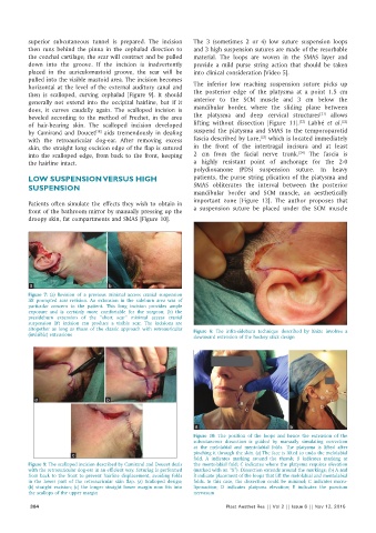
Patients often simulate the effects they wish to obtain in
front of the bathroom mirror by manually pressing up the a suspension suture be placed under the SCM muscle
droopy skin, fat compartments and SMAS [Figure 10].
a b
Figure 7: (a) Revision of a previous minimal access cranial suspension
lift prompted scar revision. An extension in the sideburn area was of
particular concern to the patient. This long incision provides ample
exposure and is certainly more comfortable for the surgeon; (b) the
presideburn extension of the “short scar” minimal access cranial
suspension lift incision can produce a visible scar. The incisions are
altogether as long as those of the classic approach with retroauricular Figure 8: The infra‑sideburn technique described by Knize involves a
(invisible) extensions downward extension of the hockey stick design
a b
a b
Figure 10: The position of the loops and hence the extension of the
subcutaneous dissection is guided by manually simulating correction
at the melolabial and mentolabial folds. The platysma is lifted after
c pinching it through the skin. (a) The face is lifted to undo the melolabial
fold. A indicates marking around the thumb; B indicates marking at
Figure 9: The scalloped incision described by Camirand and Doucet deals the mentolabial fold; C indicates where the platysma requires elevation
with the retroauricular dog‑ear in an efficient way. Suturing is performed (marked with an “X”). Dissection extends around the markings; (b) A and
from back to the front to prevent hairline displacement, avoiding folds B indicate placement of the loops that lift the melolabial and mentolabial
in the lower part of the retroauricular skin flap. (a) Scalloped design; folds. In this case, the dissection could be minimal; C indicates micro‑
(b) straight excision; (c) the longer straight lower margin now fits into liposuction; D indicates platysma elevation; E indicates the punctum
the scallops of the upper margin nervosum
304 Plast Aesthet Res || Vol 2 || Issue 6 || Nov 12, 2015