
Page 301 - Read Online
P. 301
palatoplasty technique. The patient reported for follow‑up gene, a homolog of the p53 tumor suppressor gene. The
on a regular basis for a period of six months [Figure 6]. p63 produces a protein, which is essential for ectodermal
The hypernasal quality of the voice did not improve development. The mutation is thought to affect p63 binding
following palatoplasty. Although speech therapy was to DNA, resulting in abnormal ectodermal development. [11,12]
prescribed, the patient did not return for treatment. The p63 is expressed in the ectodermal surfaces of the limb
buds. Limb truncations are due to a failure to maintain the
DISCUSSION apical ectodermal ridge, a stratified epithelium essential
for limb development. The p63 is critical for maintaining
EEC syndrome is a rare autosomal dominant multiple the progenitor‑cell populations necessary for epithelial
congenital anomaly syndromes with variable expression. development and morphogenesis. [13]
Disease‑causing mutations have been identified in the p63
There are three types of EEC syndrome, with gene loci
identified as follows:
• EEC syndrome type 1 (Mendelian Inheritance in Man (MIM)
129900)‑7q11.2‑q21.3;
• EEC syndrome type 2 (MIM 602077)‑chromosome 19;
• EEC syndrome type 3 (MIM 604292)‑3q27.
Different combinations of ectodermal dysplasia, orofacial
clefting and limb malformation are seen in five different
syndromes: EEC syndrome (most common, OMIM 604292),
ankyloblepharon‑ectodermal defect‑cleft lip/palate
syndrome (AEC, OMIM 106260), limb mammary syndrome
(LMS, OMIM 603543), acro‑dermato‑ungual‑lacrimal‑tooth
syndrome (adult, OMIM 103285), and Rapp‑Hodgkin
[14]
syndrome (RHS, OMIM 129400). The term ectrodactyly
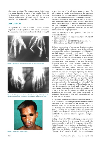
Figure 4: Radiograph of upper extremities showing ectrodactyly originates from the Greek “ektroma” (abortion) and
“daktylos” (finger). In 1829, von Walter described the
characteristic crab foot, which Cruveilhier later coined
the “lobster foot”. Ectrodactyly, cleft foot, lobster foot
[1]
and crab‑claw foot are synonyms expressing the absence
of, to a greater or lesser degree, the central digits
and metacarpals or metatarsals of the feet and hands,
respectively. The first and fifth rays are generally present,
although sometimes the first is hypertrophied. The usual
presentation involves the hands and feet bilaterally. Our
[1]
case demonstrated ectrodactyly of both hands and the
right foot. According to Blauth and Borisch six level
[15]
radiographic classification of cleft feet, the right foot is
Grade II, as there are five metatarsals, which are partially
hypoplastic or synostotic with other metatarsals or
Figure 5: Radiograph of lower extremity showing deformity of the phalanges. The second or third ray is always affected, and
second metatarsal and the absence of phalanges of the second digit at least one toe is absent.
ED syndrome requires involvement from the birth of hair,
skin, teeth or nails without progression of the condition.
The signs of ED were seen in the present case as the
hair was brittle but not sparse. The anodontia noted in
the maxillary arch may have been secondary to the cleft
palate, however, the anodontia of the mandibular arch
could be attributed to ED. The toenails were ridged, thick
and brittle. The patient’s skin was pigmented and prone
to rashes.
Making a diagnosis of ED is challenging as any ectodermal
derivative may be involved in varying degrees, and other
systems may also be affected. To circumvent this problem,
Freire‑Maia and Pinheiro designed a classification system,
which contains over 100 clinically distinct conditions. [9,15,16]
Freire‑Maia and Pinheiro based their definition on four
Figure 6: Six months postoperative “classical signs” associated with ED: trichodysplasia,
292 Plast Aesthet Res || Vol 2 || Issue 5 || Sep 15, 2015