
Page 296 - Read Online
P. 296
and the superficial fascia whereas the deep fat occupies Calipers (series‑sc02, Guilin Gunglv measuring instrument
the area between the superficial fascia and the muscles. Co. Ltd., Guilin, China); and the average readings of 3 larger
Superficial layer is richly vascularized and results in cellulite lobules was calculated for further analysis.
formation when it is hypertrophied. The deep layer of fat is Statistical analysis was performed using the SPSS 15
called localized fat deposit (LFD) when it is hypertrophied. package (SPSS, IBM Company). Data were expressed
LFD is excessive bulge producing contour deformity of
the region. The subcutaneous fat deposits in the lower as mean ± standard deviation (SD) and 95% confidence
[11]
abdomen (LA) do not get absorbed easily by dieting and interval. Paired sample t‑test was applied for comparing
exercise, compared to the upper abdomen (UA). UA and LA parameters in each sex. Independent sample
t‑test was applied for comparing the parameters between
The present study was carried to find the difference in males and females. In addition, Pearson’s correlation test
morphology of subcutaneous fat lobules, as an initial step was applied to correlate the parameters of the upper and
to explore the different re‑absorption pattern of deposited LA. P < 0.05 was considered as statistically significant.
fat different location of abdomen and different gender.
RESULTS
METHODS
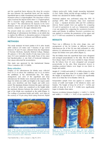
There was a difference in the color, shape, size, and
This study included 40 fresh (within 6‑10 h after death) arrangement of the fat lobules at different locations.
adult cadavers (33 males and 7 females) of age varied Subcutaneous fat of the UA was dark yellowish in color
between 18 and 70 years collected from the Department whereas LA subcutaneous fat was yellowish in color, and
of Forensic Medicine, Kasturba Medical College, Manipal. deeper fat lobules were pale yellow [Figure 2].
All cadavers (<10 h after death) of body mass index more
than 19.5 were included in this study. None of the cases The fat lobules from the superficial layer were elongated
had lipodistrophies or any kind of hormonal imbalance and arranged perpendicular to the skin. The fat lobules
that causes abnormal fat accumulation. from deeper layers of UA were rounded in shape whereas
the fat lobules from LA were elongated and arranged
This study was approved by the Institutional Human perpendicular to the skin. Most dependent (lowest in
Ethics Committee (IEC 111/2009).
standing position) lobules were larger in size (both in
Data collection height and width).
Samples of the subcutaneous fat lobules were collected Height (P = 0.042) and width (P = 0.008) of deep fat of LA
from UA (3 cm above the umbilicus) and LA (3 cm below were significantly more than UA in males [Table 1] while
the umbilicus) at the mid‑clavicular line. The shape, the height of superficial fat (P = 0.016) was significantly
arrangement, and color of the superficial and deep more in LA than the UA in females [Table 2].
subcutaneous fat lobules were observed and recorded. The
larger fat lobules (in width and height) in the region under Independent sample t‑test was applied to compare the
study were identified, and their height and width were means of width and height of fat lobules‑in males and
noted. The maximum distance between the upper and lower females. The height of deep fat of UA (P = 0.018) and
end of the fat lobule was considered as the height while width of deep fat of LA (P = 0.020) were significantly
the maximum distance between the anterior and posterior larger in females than males.
part of the lobule was considered as the width [Figure 1].
The readings of maximum height and width were taken at Pearson correlation between height (r = 0.403, P = 0.010)
the accuracy of 10 μm using metal casing Electronic Digital and width (r = 0.585, P < 0.01) of the superficial fat of
Figure 1: The schematic representation of the measurements of a fat Figure 2: The subcutaneous fat of upper and lower abdomen.
lobule. Height (Ht): the maximum distance between the upper and lower Subcutaneous fat of the UA was dark yellowish in color whereas in
end of the fat lobule; Width (Wt): the maximum distance between the the LA, subcutaneous fat was yellowish in color. UA: Upper abdomen,
anterior and posterior part of the lobule LA: Lower abdomen
Plast Aesthet Res || Vol 2 || Issue 5 || Sep 15, 2015 287