
Page 304 - Read Online
P. 304
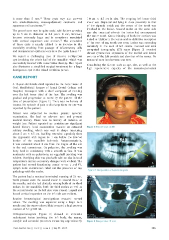
is more than 5  mm.  These cysts may also convert   3.0  cm  ×  4.5  cm in size.  The erupting left lower third
                             [1]
          into ameloblastomas, mucoepidermoid carcinoma and   molar was displaced and lying in close proximity  to that
          squamous cell carcinoma. [10]                       of the  sigmoid notch and the crown of the  tooth was
                                                              involved in  the  lesion.  Second molar  on  the  same  side
          The growth rate may be quite rapid, with lesions growing   was also impacted wherein the lesion had encompassed
          up to 5  cm  in  diameter  in  3‑4  years.  It  can, however,   the entire tooth. Gross thinning of both the cortices was
          become extremely  large and is sometimes  associated   noted in relation to the lesion and no definitive resorption
          with cortical expansion and erosion.  The expansion   of the root of any tooth was seen. Lesion was extending
                                           [3]
          of these cysts is  usually related to an increase  in the   anteriorly  to  the  root of left  canine.  Coronal and axial
          osmolality resulting from passage of inflammatory cells   computed tomography  (CT)  scans  [Figure  3] revealed
          and desquamated epithelial cells into the cystic lumen. [11]
                                                              almost  symmetrical  expansion  of the  medial  and lateral
          We  report a challenging  case  of massive  dentigerous   cortices of the left condyle and also that of the ramus. No
          cyst involving the whole half of the mandible, which was   temporal bone involvement was seen.
          successfully treated with conservative therapy. This report   Considering  the  factors such as  age,  site,  as  well as  the
          also illustrates a simplified surgical treatment  for a large   high  regenerative  capacity of the  musculo‑periosteal
          dentigerous cyst in the mixed dentition period.

          CASE REPORT

          A 13‑year‑old female child reported to the Department of
          Oral,  Maxillofacial Surgery  of Bapuji Dental College  and
          Hospital, Davangere  with  a chief complaint of swelling
          over the  left  lower third of the  face.  The  swelling  was
          gradual and progressive  as  noted by  the  patient  till  the
          time  of presentation [Figure 1]. There was no history of
          trauma. No episode of pain or discharge from the site was
          reported by the patient.
          Patient was subjected to routine general systemic
          examination.  She had no relevant past and present
          medical history.  There  was no history  of cachexia or
          weight  loss.  Patient  reported no  contributory  significant
          dental history. Local examination revealed an extraoral   Figure 1: Preoperative profile
          solitary swelling, which was oval in shape measuring
          about 5 cm × 4.5 cm. Swelling extended superiorly from
          the zygomatic arch region to 1  cm below the inferior
          border of the mandible inferiorly. Anteroposteriorly,
          it was extended about 3  cm from the tragus of the ear
          to the oral commissure. On palpation, the swelling was
          bony hard in  consistency  with a smooth surface. It  was
          nontender with no pulsations; no egg‑shell crackling was
          evident. Overlying skin was pinchable with no rise in local
          temperature and no secondary changes were evident. The
          patient had normal functioning cranial  nerves V and  VII.
          Lymph node examination  ruled out the presence of any
          pathology with the nodes.                            Figure 2: Preoperative orthopantomogram

          The patient had a maximal interincisal opening of 35 mm.
          Teeth present were the second molar to second molar in
          the maxilla, and she had clinically missing both of the third
          molars. In the mandible, both the third molars as well as
          the second molar on the left side were absent. Lingual and
          buccal cortical expansion on the left side was evident.
          Routine hematological  investigations revealed normal
          values.  The swelling  was aspirated using  a  large  bore
          needle and the straw‑colored fluid revealed a high protein
          content of 5.1 g/100 mL.
          Orthopantomogram  [Figure  2] showed an  expansile
          radiolucent lesion involving  the  left body,  the  ramus,
          condyle and coronoid processes measuring approximately   Figure 3: Preoperative CT scan

          Plast Aesthet Res || Vol 2 || Issue 5 || Sep 15, 2015                                             295