Page 305 - Read Online
P. 305
capsule of the growing child, it was planned to treat the   8th week, IMF was released. As per our protocol, CT scans
          lesion with simple enucleation procedure.           (both axial and coronal view) and radiographs were taken
                                                              at  regular  intervals  and  evaluated.  Radiographic  follow‑up
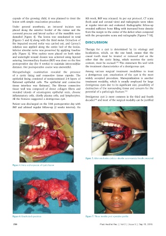
          Under  general  anesthesia,  an  intraoral  incision  was
          placed along the anterior border of the ramus and the   revealed sufficient bone filling with increased bone density
          coronoid process and lateral surface of the mandible were   from the margin to the center of the defect when compared
          denuded  [Figure  4].  The  lesion  was  enucleated  in  total   with the preoperative scans and radiographs [Figures 7‑10].
          [Figures 5 and 6] along with the third molar. Extraction of
          the impacted second molar was carried out, and Carnoy’s   DISCUSSION
          solution  was  applied  along  the  entire  bed  of  the  lesion.
          Inferior alveolar nerve was preserved by applying Vaseline   Therapy for a  cyst  is  determined  by  its  etiology  and
          jelly [Figure 5]. Wire eyelets were placed on both sides   localization, which, on the one hand, means that the
          and watertight wound closure was achieved using layered   causal tooth must be treated or removed and on the
          suturing. Intermaxillary fixation (IMF) was done on the first   other  that  the  cystic lining,  which secretes  the  cystic
                                                                                    [12]
          postoperative day (for 8 weeks) to maintain intercondylar   content, must be excised.  This statement fits well with
          distance. Her postoperative course was uneventful.  the treatment characteristics of a dentigerous cyst.
          Histopathologic examination  revealed the  presence   Among  various  surgical  treatment  modalities  to  treat
          of  a  cystic  lining  and connective  tissue  capsule. The   a  dentigerous  cyst,  enucleation  of the  cyst  is  the  most
          epithelial lining consisted of nonkeratinized 2‑4 layers of   widely accepted procedure. Marsupialization is  another
          flattened epithelial cells. The epithelial and connective   treatment modality, which is usually employed for large
          tissue  interface was flattened.  The fibrous connective   dentigerous cysts due to its significant size, possibility of
          tissue wall was composed  of dense collagen fibers and   destruction of the surrounding tissue and concern for the
          revealed islands of odontogenic epithelial rests, chronic   potential of a pathologic fracture. [13]
          inflammatory cells, chiefly plasma cells, and lymphocytes.   Dentigerous  cyst  is  more  common  in  the  third  and  fourth
          All the features suggested a dentigerous cyst.      decades  and most of the surgical modality can be justified
                                                                     [14]
          Patient was discharged on the 10th postoperative day with
          IMF and advised regular follow‑up (2  weeks interval). On


                                                              Figure 5: After enucleation inferior alveolar neurovascular bundle preserved

          Figure 4: Intra oral exposure of cystic lesion


          Figure 6: Enucleated specimen                       Figure 7: Three months post operative profile

           296                                                           Plast Aesthet Res || Vol 2 || Issue 5 || Sep 15, 2015

