
Page 284 - Read Online
P. 284
A B
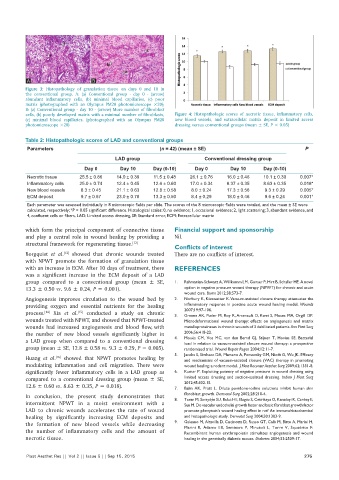
Figure 3: Histopathology of granulation tissue on days 0 and 10 in
the conventional group. A: (a) Conventional group ‑ day 0 ‑ (arrow)
abundant inflammatory cells, (b) minimal blood capillaries, (c) poor
matrix (photographed with an Olympus PM20 photomicroscope ×20);
B: (a) Conventional group ‑ day 10 ‑ (arrow) More number of fibroblast
cells, (b) poorly developed matrix with a minimal number of fibroblasts, Figure 4: Histopathologic scores of necrotic tissue, inflammatory cells,
(c) minimal blood capillaries. (photographed with an Olympus PM20 new blood vessels, and extracellular matrix deposit in limited access
photomicroscope ×20) dressing versus conventional groups (mean ± SE, P < 0.05)
Table 2: Histopathologic scores of LAD and conventional groups
Parameters (n = 42) (mean ± SE) P
LAD group Conventional dressing group
Day 0 Day 10 Day (0‑10) Day 0 Day 10 Day (0‑10)
Necrotic tissue 25.5 ± 0.86 14.0 ± 0.39 11.5 ± 0.48 26.1 ± 0.76 16.0 ± 0.46 10.1 ± 0.30 0.007*
Inflammatory cells 25.0 ± 0.74 12.4 ± 0.45 12.6 ± 0.60 17.0 ± 0.34 8.37 ± 0.35 8.63 ± 0.35 0.018*
New blood vessels 8.3 ± 0.45 21.1 ± 0.63 12.8 ± 0.58 8.0 ± 0.24 17.3 ± 0.56 9.3 ± 0.29 0.005*
ECM deposit 9.7 ± 0.67 23.0 ± 0.78 13.3 ± 0.50 8.4 ± 0.29 18.0 ± 0.46 9.6 ± 0.24 0.001*
Each parameter was assessed individually in 8 microscopic fields per slide. The scores of the 8 microscopic fields were totaled, and the mean ± SE were
calculated, respectively; *P < 0.05 significant difference. Histological scales: 0, no evidence; 1, occasional evidence; 2, light scattering; 3, abundant evidence, and
4, confluent cells or fibers. LAD: Limited access dressing, SE: Standard error, ECM: Extracellular matrix
which form the principal component of connective tissue Financial support and sponsorship
and play a central role in wound healing by providing a Nil.
structural framework for regenerating tissue. [12]
Conflicts of interest
Borgquist et al. showed that chronic wounds treated There are no conflicts of interest.
[13]
with NPWT promote the formation of granulation tissue
with an increase in ECM. After 10 days of treatment, there REFERENCES
was a significant increase in the ECM deposit of a LAD
group compared to a conventional group (mean ± SE, 1. Rahmanian‑Schwarz A, Willkomm LM, Gonser P, Hirt B, Schaller HE. A novel
13.3 ± 0.50 vs. 9.6 ± 0.24, P = 0.001). option in negative pressure wound therapy (NPWT) for chronic and acute
wound care. Burns 2012;38:573‑7.
Angiogenesis improves circulation to the wound bed by 2. Norbury K, Kieswetter K. Vacuum‑assisted closure therapy attenuates the
providing oxygen and essential nutrients for the healing inflammatory response in porcine acute wound healing model. Wounds
[15]
process. Xia et al. conducted a study on chronic 3. 2007;19:97‑106.
[14]
Greene AK, Puder M, Roy R, Arsenault D, Kwei S, Moses MA, Orgill DP.
wounds treated with NPWT, and showed that NPWT‑treated Microdeformational wound therapy: effects on angiogenesis and matrix
wounds had increased angiogenesis and blood flow, with metalloproteinases in chronic wounds of 3 debilitated patients. Ann Plast Surg
the number of new blood vessels significantly higher in 2006;56:418‑22.
a LAD group when compared to a conventional dressing 4. Mouës CM, Vos MC, van den Bemd GJ, Stijnen T, Hovius SE. Bacterial
load in relation to vacuum‑assisted closure wound therapy: a prospective
group (mean ± SE, 12.8 ± 0.58 vs. 9.3 ± 0.29, P = 0.005). randomized trial. Wound Repair Regen 2004;12:11‑7.
5. Jacobs S, Simhaee DA, Marsano A, Fomovsky GM, Niedt G, Wu JK. Efficacy
[16]
Huang et al. showed that NPWT promotes healing by and mechanisms of vacuum‑assisted closure (VAC) therapy in promoting
modulating inflammation and cell migration. There were wound healing: a rodent model. J Plast Reconstr Aesthet Surg 2009;62:1331‑8.
significantly fewer inflammatory cells in a LAD group as 6. Kumar P. Exploiting potency of negative pressure in wound dressing using
compared to a conventional dressing group (mean ± SE, limited access dressing and suction‑assisted dressing. Indian J Plast Surg
12.6 ± 0.60 vs. 8.63 ± 0.35, P = 0.018). 7. 2012;45:302‑15.
Balin AK, Pratt L. Dilute povidone‑iodine solutions inhibit human skin
In conclusion, the present study demonstrates that fibroblast growth. Dermatol Surg 2002;28:210‑4.
intermittent NPWT in a moist environment with a 8. Turan M, Saraydýn SU, Bulut HE, Elagöz S, Cetinkaya O, Karadayi K, Canbay E,
Sen M. Do vascular endothelial growth factor and basic fibroblast growth factor
LAD to chronic wounds accelerates the rate of wound promote phenytoin’s wound healing effect in rat? An immunohistochemical
healing by significantly increasing ECM deposits and and histopathologic study. Dermatol Surg 2004;30:1303‑9.
the formation of new blood vessels while decreasing 9. Galeano M, Altavilla D, Cucinotta D, Russo GT, Calò M, Bitto A, Marini H,
the number of inflammatory cells and the amount of Marini R, Adamo EB, Seminara P, Minutoli L, Torre V, Squadrito F.
Recombinant human erythropoietin stimulates angiogenesis and wound
necrotic tissue. healing in the genetically diabetic mouse. Diabetes 2004;53:2509‑17.
Plast Aesthet Res || Vol 2 || Issue 5 || Sep 15, 2015 275