
Page 283 - Read Online
P. 283
Wounds were washed daily both the LAD group Statistical analysis
and the conventional group prior to dressing with Statistical analysis was performed using the student’s t‑test
5% povidone‑iodine solution. Of these 140 patients, for comparisons between groups (SPSS, 15th version (233
56 participants (22 in the LAD group and 34 in the South Wacker Drive, 11th Floor, Chicago)). The data were
conventional dressing group) were lost to follow‑up. Of expressed as mean ± standard error (SE). P < 0.05 was
42 patients in the LAD group, 22 (52%) were women, considered to be significant. When appropriate, statistical
and 20 (48%) were men. In the conventional dressing uncertainty was expressed with 95% confidence levels.
group, 18 (42.8%) were women, and 24 (57.1%) were
men. Biopsies were taken on days 0 and 10 and were RESULTS
analyzed for the histopathologic parameters under
study. On day 0, both the LAD and conventional groups showed
Randomization necrotic tissue with increased inflammatory infiltrates
Patients were randomized by generating tables of random [Figures 2A and 3A]. On day 10, the LAD group [Figure 2B]
numbers through www.random.org. Numbers were showed an increase in ECM deposition and angiogenesis
assigned to a treatment group and sealed in opaque with a decrease in inflammatory infiltrate when
envelopes containing labeled paper with the treatment compared to the conventional group [Figure 3B]. The
and patient ID. results of the histopathologic scoring are shown in
Table 2. Histopathology revealed that in LAD group
Tissue preparation for histopathologic study after 10 days of treatment, the scores of necrotic tissue
Wound biopsies performed on days 0 and 10 were (P = 0.007) and inflammatory cell infiltrate (P = 0.018)
collected, fixed in 10% formalin, dehydrated through were significantly lower than those of the conventional
an increasing alcohol series (50%, 70%, 90%, and 100%), treatment group. The score of ECM deposits and number
cleared in xylene and embedded (Leica EG1150 H) in of blood vessels on day 0 were not well‑defined in either
paraffin wax (melting point 56°C). Serial sections of 5 µm group, but on day 10 ECM deposits (P = 0.001) and
thickness were cut using a microtome (Leica RM2255) number of blood vessel (P = 0.005) were significantly
and were stained with hematoxylin‑eosin (Sigma‑Aldrich, higher in the LAD group than in the conventional group
MO, USA). Each section was evaluated in 8 microscopic [Table 2, Figure 4].
fields (×100) under light microscopy (Olympus PM20).
Histopathology slides were graded using a modified DISCUSSION
0‑4 Ehrlich and Hunt numerical scale, and modified and
internally validated in our laboratory on a scale of 1‑3, Wound healing is a complex and dynamic process which
with 1 representing necrosis, 2 representing inflammatory involves cell‑cell interactions and cell‑matrix interaction.
cell infiltration (white blood cell and fibroblast count), and The proliferative phase of wound healing is marked by
3 representing ECM deposition. We used a 5‑point scale angiogenesis, collagen deposition, granulation tissue
to evaluate the presence of necrosis and inflammatory formation, epithelialization, and wound contraction
cell infiltration (0, no evidence; 1, occasional evidence; resulting in less scar tissue. [10]
2, light scattering; 3, abundant evidence; 4, confluent cell) A study by Nain et al. showed a decrease in the
[11]
as previously described, and used a 4‑point scale to amount of necrotic tissue in chronic wounds treated with
[8]
evaluate the presence of ECM deposition (0, no evidence; NPWT. Histopathological analysis showed significantly
1, little ECM deposition; 2, moderate ECM deposition; less necrotic tissue in the LAD group compared to a
3, confluent ECM deposition) as previously described. conventional dressing group after 10 days of treatment
[8]
In determining the degree of angiogenesis, only mature (mean ± SE, 11.5 ± 0.48 vs. 10.1 ± 0.30, P = 0.007). The
vessels were counted and identified by the presence of ECM is made up of collagen fibers and glycosaminoglycans,
erythrocytes in their lumen. The score was assigned by 2
[9]
investigators. The code describing the specific treatment
received by the patient was broken after scoring had been
completed by the investigators.
Table 1: Patient demographics and baseline wound
characterization
Details LAD group Conventional
group
A B
Number of patients 42 42
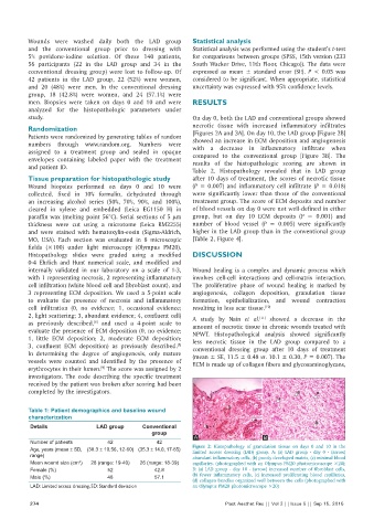
Age, years (mean ± SD, (38.3 ± 10.56, 12-60) (35.3 ± 14.0, 17-65) Figure 2: Histopathology of granulation tissue on days 0 and 10 in the
limited access dressing (LAD) group. A: (a) LAD group ‑ day 0 ‑ (arrow)
range) abundant inflammatory cells, (b) poorly developed matrix, (c) minimal blood
Mean wound size (cm ) 28 (range: 19-40) 26 (range: 18-39) capillaries. (photographed with an Olympus PM20 photomicroscope ×20);
2
Female (%) 52 42.8 B: (a) LAD group ‑ day 10 ‑ (arrow) increased number of fibroblast cells,
Male (%) 48 57.1 (b) fewer inflammatory cells, (c) increased proliferating blood capillaries,
(d) collagen bundles organized well between the cells (photographed with
LAD: Limited access dressing, SD: Standard deviation an Olympus PM20 photomicroscope ×20)
274 Plast Aesthet Res || Vol 2 || Issue 5 || Sep 15, 2015