
Page 254 - Read Online
P. 254
effects of VEGF and bFGF. Expression of the inducible 140 amino acids. Acidic FGF and bFGF are the first few
[26]
COX‑2 enzyme during the inflammatory stage of healing to be discovered and are now designated as FGF‑1 and
also leads to VEGF production and other promoters of FGF‑2, respectively. Both are preferentially involved
[42]
angiogenesis. [27] in the process of angiogenesis. [43,44] These compounds
are polypeptides of about 18 kDa, single chained and
Step 3: Vascular proliferation nonglycosylated. They transmit their signals through
Hypoxia is an important driving force for wound
angiogenesis. Expression of gene HIF‑1α, due to hypoxic FGF receptor‑4 (FGFR‑4) high‑affinity, protein family of
gradient between injured and healthy tissue triggers transmembrane tyrosine kinases (FGFR‑1 to FGFR‑4), that
VEGF production. [24,28] VEGF is present in both wound bind to different FGFs with different affinities. The strong
tissue and exudate. [28,29] VEGF is also known as vascular interactions of FGF‑1 and FGF‑2 with glycosaminoglycans,
[45]
permeability factor since it increases permeability of such as heparin sulfate present in the ECM, makes the
capillaries. Hypoxia also leads to endothelial cell FGFs stable against thermal, proteolytic denaturation and
[30]
production of nitric oxide (NO). NO promotes vasodilation limits its diffusibility. Thus, the ECM acts as a reservoir for
and angiogenesis to improve local blood flow. [31] pro‑angiogenic factors. Most members of the FGF family
act as a broad spectrum mitogen that stimulates the
Step 4: Vascular stabilization proliferation of mesenchymal cells of mesodermal origin,
Vascular stabilization is governed by Ang‑1, tyrosine kinase as well as ectodermal and endodermal cells.
with immunoglobulin‑like and EGF‑like domains 2 (Tie‑2),
smooth muscle cells and pericytes. Production of PDGF FGF‑1 and FGF‑2 are synthesized by a variety of cell types
and recruitment of smooth muscle cells and pericytes to including inflammatory cells and dermal fibroblasts that
the newly forming vasculature are regulated by binding are involved in angiogenesis and wound healing. When
of Ang‑1 to its receptor Tie‑2 on activated endothelial liberated from ECM, they act on the endothelial cells
cells. [32‑34] A PDGF deficiency leads to poorly‑formed in a paracrine manner, or when released by endothelial
immature blood vessels. [35] cell they act in an autocrine manner promoting cell
proliferation and differentiation. During the formation of
Step 5: Angiogenesis suppression granulation tissue, FGF‑2 promotes cell migration through
Angiogenesis is suppressed at the terminal stages of surface receptors for integrins, which mediate the binding
healing. As tissue hypoxia is restored, and inflammation of endothelial cells to ECM. [44]
[36]
subsides, the level of growth factors decline in the wound. Vascular endothelial growth factor increase vaso‑permeability
Pericytes which stabilize endothelial cells secrete an by increasing the fenestration and hydraulic conductivity.
inhibitory form of activated TGF‑β that impedes vascular
proliferation. [34,37,38] A cleavage product of collagen XVIII, This allows leakage of fibrinogen and fibronectin,
endostatin, is present surrounding the VBM, and it inhibits which are essential for the formation of the provisional
[46‑48]
wound vascularity. [39,40] ECM. The ECM is produced in large quantities by
the epidermis during wound healing. Low oxygen
[49]
WOUND ANGIOGENIC STIMULATORS tension that occurs in tissue hypoxia is a major inducer
of VEGF and its receptors. Thus, cell disruption and
[51]
[50]
AND INHIBITORS hypoxia appear to be strong initial inducers of potent
angiogenesis factors at the wound site. VEGF family
A number of angiogenic stimulators have been identified currently includes VEGF‑A, VEGF‑B, VEGF‑C, VEGF‑D,
in wound sand others are likely to exist that play an VEGF‑E and placental growth factor. VEGF‑A is a
[52]
important role in the repair [Table 1]. The stimulators homodimer glycoprotein whose subunits are linked by
in wound fluids are growth factors known to increase 2 disulfide bonds. VEGF‑A is synthesized from internal
endothelial cell migration and proliferation in vitro. [41] rearrangements (“alternative splicing”) of mRNA. Thus,
The FGF comprises of 23 homologous structures that there is the production of 7 isoforms with 121 to 206
are small polypeptides with a central core containing amino acids. [53‑55] Among these, the VEGF121, VEGF165,
VEGF189 and VEGF206 are the predominant isoforms.
[56]
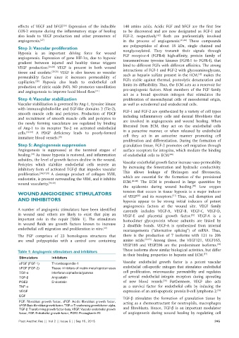
Table 1: Angiogenic stimulators and inhibitors These isoforms show similar biological activities, but differ
[57]
Stimulators Inhibitors in their binding properties to heparin and ECM.
Vascular endothelial growth factor is a potent vascular
aFGF (FGF-1) Thrombospondin-1
bFGF (FGF-2) Tissue inhibitors of matrix metalloproteinases endothelial cell‑specific mitogen that stimulates endothelial
TGF‑α Interferon alpha/beta/gamma cell proliferation, microvascular permeability and regulates
TGF‑β Angiostatin of several endothelial integrin receptors during sprouting
[58]
PGE2 Endostatin of new blood vessels. Furthermore, VEGF also acts
TNF‑α as a survival factor for endothelial cells by inducing the
VEGF expression of an anti‑apoptotic protein B‑cell lymphoma 2. [59]
EGF
TGF‑β stimulates the formation of granulation tissue by
FGF: Fibroblast growth factor, aFGF: Acidic fibroblast growth factor, acting as a chemoattractant for neutrophils, macrophages
bFGF: Basic fibroblast growth factor, TGF‑α: Transforming growth factor‑alpha,
TGF‑β: Transforming growth factor‑beta, VEGF: Vascular endothelial growth and fibroblasts. Hence, TGF‑β is an important modulator
factor, EGF: Endothelial growth factor, PGE2: Prostaglandin E2 of angiogenesis during wound healing by regulating cell
Plast Aesthet Res || Vol 2 || Issue 5 || Sep 15, 2015 245