
Page 250 - Read Online
P. 250
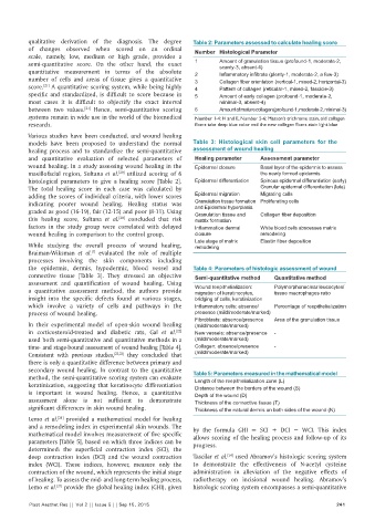
qualitative  derivation  of  the  diagnosis.  The  degree   Table 2: Parameters assessed to calculate healing score
          of changes  observed when  scored on an ordinal      Number Histological Parameter
          scale, namely,  low,  medium  or high  grade,  provides a
          semi‑quantitative  score.  On  the  other hand, the  exact   1  Amount of granulation tissue (profound-1, moderate-2,
                                                                       scanty-3, absent-4)
          quantitative  measurement in terms of the absolute   2       Inflammatory infiltrate (plenty‑1, moderate‑2, a few‑3)
          number  of cells and areas  of tissue  gives  a  quantitative   3  Collagen fiber orientation (vertical‑1, mixed‑2, horizontal‑3)
          score.  A quantitative scoring system, while being highly   4  Pattern of collagen (reticular-1, mixed-2, fascicle-3)
               [21]
          specific and standardized, is difficult to score because in   5  Amount of early collagen (profound-1, moderate-2,
          most cases it is difficult to objectify the exact interval   minimal-3, absent-4)
          between  two  values.   Hence,  semi‑quantitative  scoring   6  Amount of mature collagen (profound-1, moderate-2, minimal-3)
                            [21]
          systems remain in wide use in the world of the biomedical   Number 1‑4: H and E, Number 5‑6: Masson’s trichrome stain, old collagen
          research.                                            fibers take deep blue color and the new collagen fibers stain light blue
          Various studies have been conducted, and wound healing
          models have  been  proposed to understand the  normal   Table  3: Histological skin cell parameters for the
          healing process and to standardize the semi‑quantitative   assessment of wound healing
          and quantitative  evaluation of selected parameters of   Healing parameter  Assessment parameter
          wound healing. In a study assessing wound healing in the   Epidermal closure  Basal layer of the epidermis to assess
          maxillofacial region,  Sultana  et  al.   utilized  scoring  of  6        the newly formed epidermis
                                       [20]
          histological parameters to give a healing score  [Table  2].   Epidermal differentiation  Spinous epidermal differentiation (early)
          The total healing  score  in  each case was calculated by                 Granular epidermal differentiation (late)
          adding the scores of individual criteria, with lower scores   Epidermal migration  Migrating cells
          indicating poorer wound healing. Healing status was   Granulation tissue formation  Proliferating cells
                                                               and Epidermal hyperplasia
          graded as good (16‑19), fair (12‑15) and poor (8‑11). Using   Granulation tissue and   Collagen fiber deposition
          this healing score, Sultana  et  al.  concluded  that risk   matrix formation
                                       [20]
          factors in  the study group were correlated with delayed   Inflammation dermal   White blood cells abscesses matrix
          wound healing in comparison to the control group.    closure              remodeling
                                                               Late stage of matrix   Elastin fiber deposition
          While  studying  the overall process of wound healing,   remodeling
          Braiman‑Wiksman  et  al.   evaluated the  role  of  multiple
                              [7]
          processes involving  the  skin components including
          the epidermis, dermis, hypodermic, blood vessel and   Table 4: Parameters of histologic assessment of wound
          connective tissue [Table  3]. They stressed an objective   Semi‑quantitative method  Quantitative method
          assessment  and  quantification  of  wound healing.  Using   Wound reepithelialization:   Polymorphonuclear leucocytes/
          a quantitative  assessment  method, the  authors provide   migration of keratinocytes,   tissue macrophages ratio
          insight  into  the specific defects  found at  various stages,   bridging of cells, keratinization
          which involve a variety of cells and pathways in the   Inflammatory cells: absence/  Percentage of reepithelialization
          process of wound healing.                            presence (mild/moderate/marked)
                                                               Fibroblasts: absence/presence   Area of the granulation tissue
          In their experimental model of open‑skin wound healing   (mild/moderate/marked)
          in corticosteroid‑treated and diabetic rats, Gal  et  al.    New vessels: absence/presence   -
                                                         [22]
          used both semi‑quantitative and quantitative methods in a   (mild/moderate/marked)
          time‑ and stage‑bound assessment of wound healing [Table 4].   Collagen: absence/presence   -
          Consistent with previous studies, [22,23]  they  concluded  that   (mild/moderate/marked)
          there is only a quantitative difference between primary and
          secondary  wound  healing.  In  contrast  to  the  quantitative   Table 5: Parameters measured in the mathematical model
          method, the semi‑quantitative scoring system can evaluate   Length of the reepithelialization zone (L)
          keratinization, suggesting that keratinocyte differentiation   Distance between the borders of the wound (S)
          is important in wound healing. Hence, a quantitative   Depth of the wound (D)
          assessment alone is not sufficient to demonstrate    Thickness of the connective tissue (T)
          significant differences in skin wound healing.       Thickness of the natural dermis on both sides of the wound (N)
          Lemo et al.  provided a mathematical model for healing
                   [21]
          and a remodeling index in experimental skin wounds. The   by  the  formula  GHI  =  SCI  +  DCI  −  WCI.  This  index
          mathematical model involves measurement of five specific   allows scoring of the healing process and follow‑up of its
          parameters [Table 5], based on which three indices can be   progress.
          determined: the superficial contraction index (SCI), the
                                                                         [24]
          deep contraction index (DCI) and the wound contraction   Tascilar et al.  used Abramov’s histologic scoring system
          index  (WCI).  These  indices,  however,  measure  only  the   to demonstrate the effectiveness  of N‑acetyl cysteine
          contraction of the wound, which represents the initial stage   administration  in  alleviation  of the  negative  effects  of
          of healing. To assess the mid‑ and long‑term healing process,   radiotherapy on incisional wound healing.  Abramov’s
          Lemo et al.  provide the global healing index (GHI), given   histologic scoring system encompasses a semi‑quantitative
                   [21]
          Plast Aesthet Res || Vol 2 || Issue 5 || Sep 15, 2015                                             241