
Page 253 - Read Online
P. 253
angiogenesis is required for wound healing, its induction 3. Proteolytic enzymes released by activated endothelial cells
is beneficial in many clinical situations for achieving dissolve the basement membrane of surrounding parent
wound closure. vessels;
4. Endothelial cells proliferate and sprout outward through
PHYSIOLOGICAL CONTROL OF the basement membrane;
ANGIOGENESIS 5. Endothelial cells migrate into the wound bed using
integrins (αvβ3, αvβ5 and αvβ1) which are cell surface
Angiogenesis plays a critical role in wound healing. By adhesion molecules;
developing capillary sprouts, which digest endothelial 6. Matrix metalloproteinases (MMPs) dissolve the surrounding
cells and invade the extracellular matrix (ECM) stroma tissue matrix in the path of sprouting vessels;
after penetrating through the underlying vascular 7. Vascular sprouts form tubular channels that connect to
basement membrane (VBM), and form tube‑like structures form vascular loops;
that continue to extend, branch, and form networks. 8. Vascular loops differentiate into afferent (arterial) and
During angiogenesis capillary advancement in ECM occurs efferent (venous) limbs;
by endothelial cell proliferation and direction of growth 9. New blood vessels mature by recruiting mural cells
is guided by chemotaxis from the target region. The (smooth muscle cells and pericytes) to stabilize the
interaction among endothelial cells, angiogenesis factors vascular architecture;
and surrounding ECM proteins is temporally and spatially 10. Blood flow begins in the mature stable vessel.
synchronized. [9,10]
These complex growth factor‑receptor, cell‑cell and cell‑matrix
Angiogenesis can be induced in response to injury via interactions characterize the angiogenesis process, regardless
pro‑ and anti‑angiogenic factors present throughout the of the stimuli or its location in the body.
body. Pro‑angiogenic factors consist of thrombin, fibrinogen
fragments, thymosin‑β4 and growth factors. Angiogenic
growth factors are stored in platelets and inflammatory THE ANGIOGENESIS MODEL OF
cells that circulate in the bloodstream, and are sequestered WOUND HEALING
within the ECM. The production of these factors is
regulated by genes expressed in response to hypoxia and Wound healing occurs in four major overlapping stages:
inflammation, such as hypoxia‑inducible factors (HIF) and (1) hemostatic, (2) inflammatory stage, (3) proliferative
cyclooxygenase‑2 (COX‑2). [11‑13] In contrast, angiogenesis stage, and (4) remodeling stage. Although granulation
inhibitor factors suppress blood vessel growth. [14,15] Some is assigned to the proliferative stage, angiogenesis is
inhibitors circulate in the blood stream at low physiological initiated immediately after tissue injury and is mediated
levels while others are stored in the ECM surrounding throughout the wound healing process.
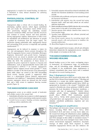
blood vessels. Vascular growth is suppressed when Step 1: Angiogenesis initiation
there is a physiological balance between angiogenesis Basic fibroblast growth factor (bFGF) stored within intact cells
stimulators and inhibitors. Immediately following injury, and the ECM is released from damaged tissue. Bleeding and
[15]
[16]
however, angiogenic stimuli are released into the wound hemostasis in a wound also initiate angiogenesis. Cellular
bed, and a shift occurs in regulators favoring vascular receptors for vascular endothelial growth factor (VEGF) are
growth [Figure 1].
upregulated by thrombin in the wound. Endothelial cells
[17]
exposed to thrombin also release gelatinase A (MMP‑2), which
THE ANGIOGENESIS CASCADE promotes the local dissolution of basement membrane, a
necessary early step of angiogenesis. Platelets release
[18]
Angiogenesis occurs as an orderly cascade of molecular multiple growth factors, including platelet‑derived
and cellular events in the wound bed: growth factor (PDGF), VEGF, transforming growth
1. Endothelial cell surface has receptors to which angiogenic factor (TGF‑α, TGF‑β), bFGF, platelet‑derived endothelial
growth factors bind in preexisting venules (parent vessels);
2. Growth factor‑receptor binding activates signaling cell growth factor and angiopoietin‑1 (Ang‑1). These factors
stimulate endothelial proliferation, migration and tube
pathways within endothelial cells;
formation. [19‑22]
Step 2: Angiogenesis amplification
Macrophages and monocytes release numerous angiogenic
factors, including PDGF, VEGF, Ang‑1, TGF‑α, bFGF,
interleukin‑8 (IL‑8) and tumor necrosis factor alpha into
the wound bed during the inflammatory phase amplifying
angiogenesis further. [23,24] Several growth factors (PDGF, VEGF
and bFGF) synergize in their ability to vascularize tissues.
[25]
Proteases that break down damaged tissue matrix further
release matrix‑bound angiogenic stimulators. Enzymatic
cleavage of fibrin yields fibrin fragment E, which
Figure 1: Angiogenesis is a balance between stimulators (growth factors)
and inhibitors as shown in this model stimulates angiogenesis directly and also enhances the
244 Plast Aesthet Res || Vol 2 || Issue 5 || Sep 15, 2015