
Page 249 - Read Online
P. 249
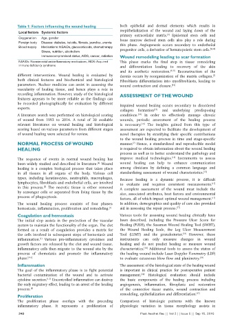
Table 1: Factors influencing the wound healing      both epithelial and dermal elements which results in
           Local factors Systemic factors                     reepithelialization of the wound and laying down of the
                                                                                      [3]
                                                              primary extracellular matrix.  Epidermal stem cells and
           Oxygenation  Age, gender                           bone marrow derived stem cells also play a role during
           Foreign body  Disease: diabetes, keloids, fibrosis, jaundice, uremia
           Blood supply  Medications: NSAIDs, glucocorticoids, chemotherapy  this phase. Angiogenesis occurs secondary to endothelial
                                                                                                            [9,10]
                      Stress, nutrition, alcoholism           progenitor cells, a derivative of hematopoietic stem cells.
                      Immunocompromised status, AIDS, cancer, radiation  Wound remodeling leading to scar formation
           NSAIDs: Nonsteroidal anti‑inflammatory medications, AIDS: Acquired   This phase marks the final step in tissue remodeling
           immune deficiency syndrome                         and differentiation  leading to recovery of the  skin
                                                              and its  aesthetic restoration. [8,11]  Reconstruction of the
          different  interventions.  Wound healing  is  evaluated by   dermis occurs by reorganization of the matrix collagen.
                                                                                                              [7]
          both clinical features  and biochemical and histological   Fibroblasts differentiation into myofibroblasts, leading to
          parameters.  Nuclear medicine  can assist  in assessing  the   wound contraction and closure. [12]
          vascularity of healing tissue, and hence plays a role in
          recording inflammation. However, study of the histological   ASSESSMENT OF THE WOUND
          features appears to be more reliable as the findings can
          be  recorded photographically for evaluation by  different   Impaired wound healing  occurs  secondary  to disordered
          experts.
                                                              collagen formation   and underlying  predisposing
                                                                                [13]
          A literature search was performed on histological scoring   conditions.   In  order to  effectively  manage  chronic
                                                                       [14]
          of wound from 1993  to 2014. A  total of 30  available   wounds, periodic assessment of the  healing  process
          relevant literatures on wound healing and  histological   is  necessary.  The insights  gained  from  this  type  of
                                                                         [15]
          scoring based on various parameters from different stages   assessment are expected to facilitate the development of
          of wound healing were selected for review.          novel therapies by  stratifying  their  specific contributions
                                                              to the wound healing process in time and stage‑specific
          NORMAL PROCESS OF WOUND                             manner.  Hence, a standardized and reproducible model
                                                                     [7]
          HEALING                                             is required to obtain information about the wound healing
                                                              process as well as to better understand the pathology and
                                                                                         [16]
          The  sequence  of events  in  normal  wound healing  has   improve medical technologies.  Instruments to assess
                                                   [4]
          been widely studied and described in literature.  Wound   wound  healing can help to enhance communication
          healing  is a complex biological process that takes place   among clinicians by defining a common language and
          in all tissues  in all organs of the body. Various cell   standardizing assessment of wound characteristics. [15]
          types,  including keratinocytes, neutrophils, macrophages,   Because  healing  is  a  dynamic  process,  it  is  difficult
          lymphocytes, fibroblasts and endothelial cells, are involved   to  evaluate  and requires  consistent  measurements.
                                                                                                             [17]
          in this process.  The necrotic tissue  is  either removed   A complete assessment of the wound must include the
                       [3]
          by scavenger cells or separated from living tissue  by the   size, associated attributes, host factors and environmental
          process of phagocytosis.                            factors, all of which impact optimal wound management.
                                                                                                             [17]
          The wound healing process consists of four phases:   In addition, demographics and quality of care also provided
          hemostasis, inflammation, proliferation and remodeling. [1]  aid in assessing the repair process. [17]
          Coagulation and hemostasis                          Various tools for assessing  wound healing  clinically have
          The initial step assists in the protection of the vascular   been  described,  including  the  Pressure  Ulcer Score  for
          system to maintain the functionality of the organ. The clot   Healing (PUSH), the Sussman Wound Healing Tool (SWHT),
          formed as a result of coagulation provides a matrix for   the Wound Healing Scale, the Leg  Ulcer Measurement
                                                                                              [18]
          the cells involved in subsequent steps of hemostasis and   Tool (LUMT) and the  granulometer.  However,  these
          inflammation.  Various pro‑inflammatory cytokines and   instruments  can only measure changes in wound
                     [1]
          growth factors are released by the clot and wound tissue.   healing and  do not predict healing or measure wound
          Inflammatory cells then migrate to the wound site by the   characteristics.  Additional  tools to assess the status of
                                                                           [18]
          process of chemotaxis and promote the inflammatory   the healing wound include Laser‑Doppler Flowmetry (LDF)
          phase. [4‑6]                                        to evaluate cutaneous blow flow and planimetry. [19]
          Inflammation                                        The assessment of the histological state of the healing wound
          The goal of the inflammatory  phase is  to fight  potential   is important in clinical practice for postoperative patient
          bacterial contamination of the wound and to activate   management.  Histological evaluation should include
                                                                         [20]
          cytokine secretion.  Uncontrolled inflammation can destroy   the basic  components of the healing  process  including
                         [1,7]
          the early migratory effect, leading to an arrest of the healing   angiogenesis, inflammation, fibroplasia and restoration
          process. [8]                                        of the connective tissue matrix, wound contraction and
                                                              remodeling, epithelialization and differentiation. [17]
          Proliferation
          The proliferation phase overlaps with the preceding   Comparison  of histologic  patterns  with  the  known
          inflammatory phase. It represents a proliferation of   physiologic variation in tissue morphology assists in
           240                                                           Plast Aesthet Res || Vol 2 || Issue 5 || Sep 15, 2015