
Page 151 - Read Online
P. 151
maxillectomy and was subsequently referred to the
Department of Prosthodontics and Implantology, Eklavya
Dental College and Hospital, Rajasthan, India. Immediate
surgical reconstruction was not recommended given the
need for further treatment with radiation therapy. External
beam postoperative radiotherapy was administered over
a period of 6 weeks. The patient tolerated the radiation
well and was subsequently referred to possible prosthetic
restoration of the oral defect after radiation therapy.
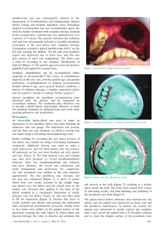
Examination revealed a partial maxillectomy defect on the
left side crossing the midline. The left side naso-maxillary
region was depressed due to bone loss, and this was
also evident in extra oral examination. The defect was
a Class IV according to the Aramany Classification of
Defects [Figure 1]. The patient agreed to have his pictures
[3]
published and signed the consent form. Figure 1: Intraoral view of the maxillectomy defect
Aesthetic rehabilitation can be accomplished either
surgically or prosthetically. The choice of rehabilitation
[4]
depends on the site, size, severity, patient age, and patient
preference. Contraindications to surgical reconstruction
include advanced age, poor general medical condition, a
history of radiation therapy, a complex anatomical defect
and the patient’s refusal to undergo further surgery. [5]
Various modalities for prosthetic reconstruction were
discussed with the patient, and he requested an
economical solution. The treatment plan therefore was
to provide a plastic-based, light-weight obturator to meet
the aesthetic demands by replacing bone and teeth while
assisting phonetics and mastication.
Procedure
An irreversible hydrocolloid was used to make an
impression of the maxillary defect area after blocking all Figure 2: Blocking of undercuts on master cast by plaster and wax
undercuts with wet gauge. The impression was poured,
and the final cast was obtained, on which a custom tray
was made using a self-curing autopolymerising resin.
Border molding for recording the soft tissue borders of
the defect was carried out using a low-fusing impression
compound. Additional silicone was used to make a
wash impression, and the final master cast was poured.
All undercuts on the cast were blocked out with plaster
and wax [Figure 2]. The final denture base and occlusal
wax rims were prepared to record maxillomandibular
relations. After the maxillomandibular jaw relations
had been obtained, the record was articulated, and
teeth arrangement was performed. On completion,
the wax prosthesis was verified at the trial insertion
appointment. The wax prosthesis was invested, and
the wax was eliminated [Figure 3]. A sheet of plastic Figure 3: Master cast after elimination of wax
based heat cure acrylic polymer in the dough stage
was placed over the defect and the palatal area on the salt present in the bulb [Figure 7], resulting in a hollow
master cast. Pressure then applied to the base of the space inside the bulb. The holes were sealed with a layer
defect resulted in a cup-shaped depression of acrylic of self-curing acrylic, and final finishing and polishing of
polymer over the defect [Figure 4]. Salt was then used the prosthesis was done [Figure 8].
to fill the depression [Figure 5]. Another thin sheet of The plastic-based hollow obturator was inserted into the
acrylic polymer was placed, and packing was performed defect, and the patient was instructed on home care and
with conventional prosthodontic protocols. Finally, three the prosthesis maintenance. To sanitize the wound, the
to four holes were drilled on the palatal surface of the patient was instructed to gently remove any exudates
prosthesis covering the bulb [Figure 6]. Warm water was with a wet cotton tip soaked with a 5% Betadine solution
injected through the holes to dissolve and eliminate the and to clean the intaglio surface of the prosthesis once
Plast Aesthet Res || Vol 2 || Issue 3 || May 15, 2015 141