
Page 101 - Read Online
P. 101
Page 14 of 22 Vakhshori et al. Plast Aesthet Res 2023;10:36 https://dx.doi.org/10.20517/2347-9264.2022.78
Table 4. Reported outcomes of free rectus femoris transfer. MRC: Medical Research Council muscle grade
Elbow Mean
Number Mean
Reference of age Pathology Neurotization Vessel flexion MRC MRC elbow Complications
anastomosis
≥ 4
flexion
MRC < 3
patients (years)
3 (degrees)
[16
Chuang et al. 1 Not Brachial Intercostal Not specified 0 1 0 NR None reported
]
specified plexus nerves
trauma
Akasaka et al. [ 11 Not Brachial Intercostal Anterior 3 8 0 80+ in 8 2 failures,
80]
specified plexus nerves (3, 4) circumflex 100+ in 3 thrombosis
trauma humeral artery or
profunda brachii
artery; cephalic
vein or brachial
vena comitantes
Wechselberger 1 22 Brachial Spinal Brachial artery and 0 0 1 110 None reported
et al. [79] plexus accessory nerve vein
trauma
[81]
Doi et al. 7 25 Brachial Spinal Thoracoacromial NR NR NR 34 3 skin paddle
plexus accessory nerve artery; cephalic necrosis
trauma vein
Terzis et al. [44] 7 NR Brachial 4 contralateral Not specified Mean muscle grade NR None reported
plexus C7 reported
trauma 2 intercostals Intercostal 2.77
1 cervical plexus Cervical plexus 2.33
cC7 3.67
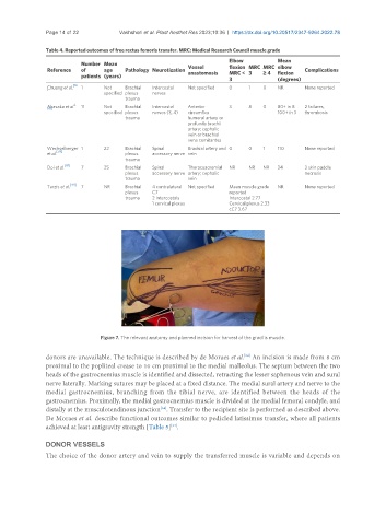
Figure 7. The relevant anatomy and planned incision for harvest of the gracilis muscle.
donors are unavailable. The technique is described by de Moraes et al. An incision is made from 8 cm
[34]
proximal to the popliteal crease to 10 cm proximal to the medial malleolus. The septum between the two
heads of the gastrocnemius muscle is identified and dissected, retracting the lesser saphenous vein and sural
nerve laterally. Marking sutures may be placed at a fixed distance. The medial sural artery and nerve to the
medial gastrocnemius, branching from the tibial nerve, are identified between the heads of the
gastrocnemius. Proximally, the medial gastrocnemius muscle is divided at the medial femoral condyle, and
distally at the musculotendinous junction . Transfer to the recipient site is performed as described above.
[34]
De Moraes et al. describe functional outcomes similar to pedicled latissimus transfer, where all patients
achieved at least antigravity strength [Table 5] .
[34]
DONOR VESSELS
The choice of the donor artery and vein to supply the transferred muscle is variable and depends on