
Page 29 - Read Online
P. 29
In the vast majority of patients, breast amyloidosis is part Comorbidities, on regular treatment, were hyperthyroidism,
of a systemic AL type disease (usually kappa light chain hypertension and heart failure.
proteins). It can be associated with malignancies of the
breast including invasive ductal or lobular carcinoma but Although the first mammogram did not suspect a
mainly it is associated with hematologic malignancies. malignant lesion, but only showed heterogeneously
dense breast, the clinical suspicion was breast cancer or
Moreover, breast cancer may sometimes be the cause of silicon leakage.
amyloid, the so-called amyloid tumour of the breast but
it is rare. [3] Magnetic resonance imaging (MRI) did not show implant
rupture. A fine needle aspiration cytology was performed,
The typical clinical presentation of breast amyloidosis is which was negative for malignancy, and reported a non-
a painless, solitary or multiple breast mass. Mammogram specific inflammatory reaction only.
shows a mass of focal or diffuse density with or without
calcification. At 2 years follow-up, the mass size increased to 3 cm. An
ultrasound guided core needle biopsy was performed
and the histological examination showed amyloid
CASE REPORT
deposits but no evidence of cancer. Amyloid deposits
appeared as eosinophilic amorphous material with
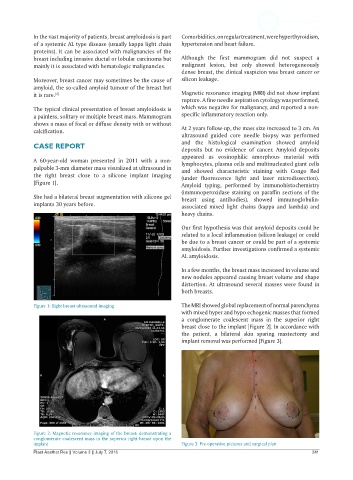
A 60-year-old woman presented in 2011 with a non- lymphocytes, plasma cells and multinucleated giant cells
palpable 3-mm diameter mass visualized at ultrasound in and showed characteristic staining with Congo Red
the right breast close to a silicone implant imaging (under fluorescence light and laser microdissection).
[Figure 1]. Amyloid typing, performed by immunohistochemistry
(immunoperoxidase staining on paraffin sections of the
She had a bilateral breast augmentation with silicone gel breast using antibodies), showed immunoglobulin-
implants 30 years before. associated mixed light chains (kappa and lambda) and
heavy chains.
Our first hypothesis was that amyloid deposits could be
related to a local inflammation (silicon leakage) or could
be due to a breast cancer or could be part of a systemic
amyloidosis. Further investigations confirmed a systemic
AL amyloidosis.
In a few months, the breast mass increased in volume and
new nodules appeared causing breast volume and shape
distortion. At ultrasound several masses were found in
both breasts.
Figure 1: Right breast ultrasound imaging The MRI showed global replacement of normal parenchyma
with mixed hyper and hypo echogenic masses that formed
a conglomerate coalescent mass in the superior right
breast close to the implant [Figure 2]. In accordance with
the patient, a bilateral skin sparing mastectomy and
implant removal was performed [Figure 3].
Figure 2: Magnetic resonance imaging of the breast: demonstrating a
conglomerate coalescent mass in the superior right breast upon the
implant Figure 3: Pre operative pictures and surgical plan
Plast Aesthet Res || Volume 3 || July 7, 2016 241