
Page 30 - Read Online
P. 30
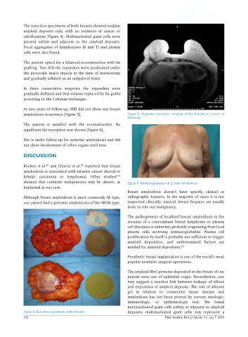
The resection specimens of both breasts showed nodular
            amyloid deposits only, with no evidence of cancer or
            calcifications [Figure 4]. Multinucleated giant cells were
            present  within  and adjacent to the  amyloid  deposits.
            Focal aggregates of lymphocytes (B and T) and plasma
            cells were also found.

            The patient opted for a bilateral reconstruction with fat
            grafting. Two 470 mL expanders were positioned under
            the pectoralis major muscle at the time of mastectomy
            and gradually inflated on an outpatient basis.

            In three consecutive surgeries  the expanders were
            gradually deflated and that volume replaced by fat grafts
            according to the Coleman technique.

            At two years of follow-up, MRI did not show any breast
            amyloidosis recurrence [Figure 5].                Figure 5: Magnetic resonance imaging of the breasts at 2 years of
                                                              follow-up
            The patient is  satisfied  with the reconstruction. No
            significant fat resorption was shown [Figure 6].

            She is under follow-up for systemic amyloidosis and did
            not show involvement of other organs until now.

            DISCUSSION

                                        [5]
            Röcken et al.  and Charlot et al.  reported that breast
                       [4]
            amyloidosis is associated with invasive cancer (ductal or
            lobular carcinoma or lymphoma). Other studies [6,7]
            showed that  comitant  malignancies  may  be  absent,  as   Figure 6: Patient appearance at 2 years of follow-up
            happened in our case.
                                                              Breast amyloidosis doesn’t have specific clinical or
            Although breast amyloidosis is most commonly AL type,   radiographic features. In the majority of cases it is not
            our patient had a systemic amyloidosis of the AH/AL type.   suspected clinically; instead, breast biopsies are usually
                                                              done to rule out malignancy.

                                                              The pathogenesis of localized breast amyloidosis in the
                                                              absence of a concomitant breast lymphoma or plasma
                                                              cell dyscrasia is unknown, probably originating from local
                                                              plasma cells secreting  immunoglobulins.  Plasma  cell
                                                              proliferation by itself is probably not sufficient to trigger
                                                              amyloid deposition, and undetermined  factors are
                                                              needed for amyloid deposition. [5]

                                                              Prosthetic breast implantation is one of the world’s most
                                                              popular aesthetic surgical operations.

                                                              The amyloid fibril proteins deposited in the breast of our
                                                              patient were not of epithelial origin. Nevertheless, one
                                                              may suggest a reaction link between leakage of silicon
                                                              and deposition of amyloid deposits. The role of silicone
                                                              gel in relation to connective tissue disease and
                                                              amyloidosis has not been proved by current serologic,
                                                              immunologic,  or epidemiologic test.  We found
                                                              multinucleated giant cells within or adjacent to amyloid
            Figure 4: Resection specimens of the breasts      deposits; multinucleated giant  cells may  represent  a
            242                                                                 Plast Aesthet Res || Volume 3 || July 7, 2016