
Page 69 - Read Online
P. 69
Page 4 of 11 Yu et al. Plast Aesthet Res 2022;9:37 https://dx.doi.org/10.20517/2347-9264.2021.124
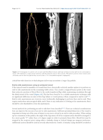
Figure 1. CT Arteriogram of a patient with an open right tibia-fibula fracture, and large soft tissue eschar and necrotic subcutaneous
tissue. CTA identified a single-vessel extremity with thrombosed anterior tibial vessels. The patent posterior tibial vessels were
ultimately used for flow-through free flap reconstruction. CTA: Computed tomographic angiography.
critical but wide dissection to find adequate inflow may necessitate a longer flap pedicle.
End-to-side anastomoses using an uninjured vessel
If the injured vessel is unusable or if vessels have been chronically occluded, another option is to perform an
end-to-side anastomosis on the remaining viable artery. This creates a surgical branch point in the vessel
that redirects a portion of blood flow to the newly transferred flap while maintaining circulation through
the distal extent of the vessel [Figure 2A]. This has been found to be a reliable anastomotic technique with
no significant difference in rates of free flap failure or vessel patency when compared to end-to-end [23-25] .
End-to-side anastomoses are, however, more technically challenging to perform than end-to-end. They
require meticulous microsurgical skills, and if there is any indication of clotting at the anastomosis, there
should be a low threshold to revise the anastomosis.
Several methods for performing an end-to-side have been described [26-28] . There are technical considerations
that can improve the success of an end-to-side anastomosis. Sufficient proximal dissection of the vessel is
important as being out of the zone of injury is even more critical in an end-to-side procedure. When setting
up the orientation of the pedicle, the angle of the flap artery off of the recipient artery should be arranged to
be a more gentle “V” rather than a 90-degree angle in order to promote linear flow. Blood flow may be
stopped in the recipient artery using either a Satinsky clamp or Acland clamps per surgeon preference.
Additional caution should be used in severely atherosclerotic vessels. A Satinsky clamp should be avoided as