
Page 72 - Read Online
P. 72
Yu et al. Plast Aesthet Res 2022;9:37 https://dx.doi.org/10.20517/2347-9264.2021.124 Page 7 of 11
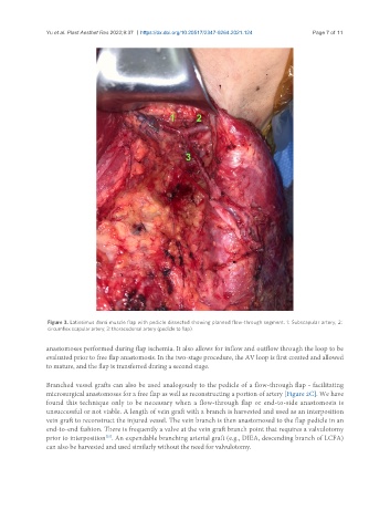
Figure 3. Latissimus dorsi muscle flap with pedicle dissected showing planned flow-through segment. 1: Subscapular artery; 2:
circumflex scapular artery; 3: thoracodorsal artery (pedicle to flap).
anastomoses performed during flap ischemia. It also allows for inflow and outflow through the loop to be
evaluated prior to free flap anastomosis. In the two-stage procedure, the AV loop is first created and allowed
to mature, and the flap is transferred during a second stage.
Branched vessel grafts can also be used analogously to the pedicle of a flow-through flap - facilitating
microsurgical anastomoses for a free flap as well as reconstructing a portion of artery [Figure 2C]. We have
found this technique only to be necessary when a flow-through flap or end-to-side anastomosis is
unsuccessful or not viable. A length of vein graft with a branch is harvested and used as an interposition
vein graft to reconstruct the injured vessel. The vein branch is then anastomosed to the flap pedicle in an
end-to-end fashion. There is frequently a valve at the vein graft branch point that requires a valvulotomy
prior to interposition . An expendable branching arterial graft (e.g., DIEA, descending branch of LCFA)
[35]
can also be harvested and used similarly without the need for valvulotomy.