
Page 73 - Read Online
P. 73
Page 8 of 11 Yu et al. Plast Aesthet Res 2022;9:37 https://dx.doi.org/10.20517/2347-9264.2021.124
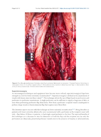
Figure 4. Flow-through anastomosis of latissimus dorsi flap to posterior tibial vessels as recipients. 1: Proximal Posterior tibial artery; 2:
subscapular artery; 3: circumflex scapular artery; 4: thoracodorsal artery (pedicle to latissimus dorsi flap); 5: distal posterior tibial
artery; 6: venous anastomosis. *Denotes arterial end-to-end anastomoses.
Supermicrosurgery
As microsurgical techniques and equipment have become more refined, supermicrosurgical flaps have
emerged as a tool for lower extremity reconstruction . Supermicrosurgery is defined as the anastomosis of
[36]
vessels with lumen sizes less than 0.8mm . This technique allows the use of flaps based on perforators and
[37]
perforator-to-perforator anastomoses. A single perforator can be sufficient to supply a large area of skin, as
seen when performing perforator flap dissections. Flow from a perforator recipient vessel is inadequate to
perfuse a large muscle or fasciocutaneous flap that requires more blood flow.
The literature reports success with this technique in lower extremity reconstruction [38,39] . Hong describes 42
patients with lower extremity defects who were reconstructed with a perforator as the recipient vessel with
only 1 flap failure. Of those 42 patients, 13 had single-vessel perfusion to the foot. The reported benefits of
this technique are a decrease in time for dissection of both the flap and the recipient site (as only the
perforators are dissected), preserved perforator vessels even in the presence of trauma or atherosclerosis,