
Page 70 - Read Online
P. 70
Yu et al. Plast Aesthet Res 2022;9:37 https://dx.doi.org/10.20517/2347-9264.2021.124 Page 5 of 11
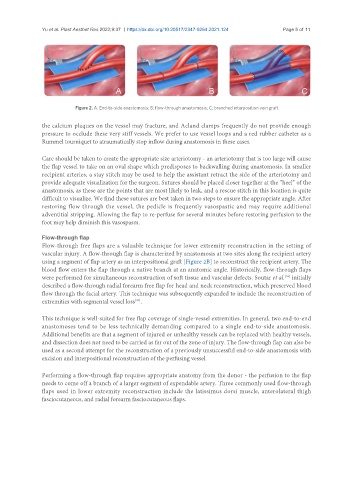
Figure 2. A: End-to-side anastomosis; B: flow-through anastomosis; C: branched interposition vein graft.
the calcium plaques on the vessel may fracture, and Acland clamps frequently do not provide enough
pressure to occlude these very stiff vessels. We prefer to use vessel loops and a red rubber catheter as a
Rummel tourniquet to atraumatically stop inflow during anastomosis in these cases.
Care should be taken to create the appropriate size arteriotomy - an arteriotomy that is too large will cause
the flap vessel to take on an oval shape which predisposes to backwalling during anastomosis. In smaller
recipient arteries, a stay stitch may be used to help the assistant retract the side of the arteriotomy and
provide adequate visualization for the surgeon. Sutures should be placed closer together at the “heel” of the
anastomosis, as these are the points that are most likely to leak, and a rescue stitch in this location is quite
difficult to visualize. We find these sutures are best taken in two steps to ensure the appropriate angle. After
restoring flow through the vessel, the pedicle is frequently vasospastic and may require additional
adventitial stripping. Allowing the flap to re-perfuse for several minutes before restoring perfusion to the
foot may help diminish this vasospasm.
Flow-through flap
Flow-through free flaps are a valuable technique for lower extremity reconstruction in the setting of
vascular injury. A flow-through flap is characterized by anastomosis at two sites along the recipient artery
using a segment of flap artery as an interpositional graft [Figure 2B] to reconstruct the recipient artery. The
blood flow enters the flap through a native branch at an anatomic angle. Historically, flow-through flaps
[29]
were performed for simultaneous reconstruction of soft tissue and vascular defects. Soutar et al. initially
described a flow-through radial forearm free flap for head and neck reconstruction, which preserved blood
flow through the facial artery. This technique was subsequently expanded to include the reconstruction of
[30]
extremities with segmental vessel loss .
This technique is well-suited for free flap coverage of single-vessel extremities. In general, two end-to-end
anastomoses tend to be less technically demanding compared to a single end-to-side anastomosis.
Additional benefits are that a segment of injured or unhealthy vessels can be replaced with healthy vessels,
and dissection does not need to be carried as far out of the zone of injury. The flow-through flap can also be
used as a second attempt for the reconstruction of a previously unsuccessful end-to-side anastomosis with
excision and interpositional reconstruction of the perfusing vessel.
Performing a flow-through flap requires appropriate anatomy from the donor - the perfusion to the flap
needs to come off a branch of a larger segment of expendable artery. Three commonly used flow-through
flaps used in lower extremity reconstruction include the latissimus dorsi muscle, anterolateral thigh
fasciocutaneous, and radial forearm fasciocutaneous flaps.