
Page 42 - Read Online
P. 42
Gimenez et al. Plast Aesthet Res 2022;9:28 https://dx.doi.org/10.20517/2347-9264.2021.129 Page 7 of 11
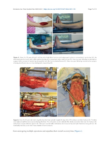
Figure 4. View of a 13-year-old girl’s left leg after high tibial fracture and subsequent anterior compartment syndrome (A). An
innervated gracilis muscle with a skin paddle was placed to reconstruct defect and function (B). View one year following reconstructive
surgery (after excision of monitoring skin paddle) with the foot in plantar flexion (C). View one-year following reconstructive surgery
showing dorsiflexion via a re-innervated gracilis muscle flap (D).
Figure 5. View of a 13-year-old male’s leg after transfer from outside hospital 10 days after ATV rollover and tibia fracture (A). The tibia
was degloved and has been exposed during this time. The orthopedic service debrided the clearly nonviable tibia near the fracture site
and placed a large antibiotic spacer (B). This was covered with a latissimus dorsi muscle flap and split-thickness skin graft (C). He
eventually had bony union after many months of bone transport.
from undergoing multiple operations and expedites their overall recovery time [Figure 6].