
Page 40 - Read Online
P. 40
Gimenez et al. Plast Aesthet Res 2022;9:28 https://dx.doi.org/10.20517/2347-9264.2021.129 Page 5 of 11
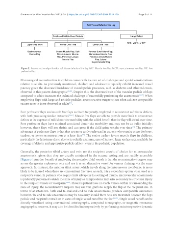
Figure 2. Reconstructive algorithm for soft tissue defects of the leg. MFF: Muscle free flap; MCFF: myocutaneous free flap; FPF: free
perforator flap.
Microsurgical reconstruction in children comes with its own set of challenges and special considerations
relative to adults. As previously mentioned, children and adolescents typically exhibit increased vessel
patency given the decreased incidence of vasculopathic processes, such as diabetes and atherosclerosis,
observed in this patient demographic [54,55] . Despite this, the decreased size of the vascular pedicle of flaps
compared to adults increases the technical challenge of successfully performing the anastomosis [56,57] . When
employing flaps with large and reliable pedicles, reconstructive surgeons can often achieve comparable
success rates to those observed in adults .
[58]
Free perforator flaps and muscle free flaps are both frequently employed to reconstruct soft tissue defects,
with both producing similar outcomes [59-61] . Muscle free flaps are able to provide more bulk to reconstruct
defects at the expense of mild donor site morbidity with the added benefit that the flap will shrink over time.
Free perforator flaps have minimal associated donor site morbidity and may not be as bulky initially;
however, these flaps will not shrink and can grow if the child gains weight over time . The primary
[62]
advantage of perforator flaps is that they are more easily reelevated in patients who require access for bony,
tendon, or nerve reconstruction at a later date . The senior author favors muscle flaps in children,
[53]
particularly the latissimus dorsi, due to its reliable anatomy, ease of harvest, large surface area available for
coverage of defects, and appropriate pedicle caliber - even in the pediatric population.
Generally, the posterior tibial artery and vein are the recipient vessels of choice for microvascular
anastomosis, given that they are usually uninjured in the trauma setting and are readily accessible
[Figure 3]. Another benefit of employing the posterior tibial vessels is that the reconstructive surgeon may
access the greater saphenous vein and use it as an alternative vessel for venous drainage via the same
approach. In contrast, the anterior tibial artery, which travels along the interosseous membrane, is more
likely to be injured when there are concomitant fractures; as such, it is a secondary option when used as a
recipient’s vessel. In patients who require limb salvage in the setting of trauma, microvascular anastomosis
is preferably performed outside the zone of injury as complications may arise secondary to structural injury
to the recipient vessels or vasospasm [63,64] . Should a patient have no viable vessels within or surrounding the
zone of injury, the reconstructive surgeon may use vein grafts to supply the flap at the recipient site. In
terms of anastomosis, both end-to-end and end-to-side anastomoses produce comparable outcomes;
however, the end-to-side anastomosis may be necessary should there be a size mismatch between the flaps
pedicle and recipient’s vessels or in cases of single-vessel runoff to the foot [53,65] . Single vessel runoff can be
directly visualized using conventional arteriography, computed tomography, or magnetic resonance
angiography; however, it may also be identified intraoperatively by performing an intraoperative dynamic