
Page 38 - Read Online
P. 38
Gimenez et al. Plast Aesthet Res 2022;9:28 https://dx.doi.org/10.20517/2347-9264.2021.129 Page 3 of 11
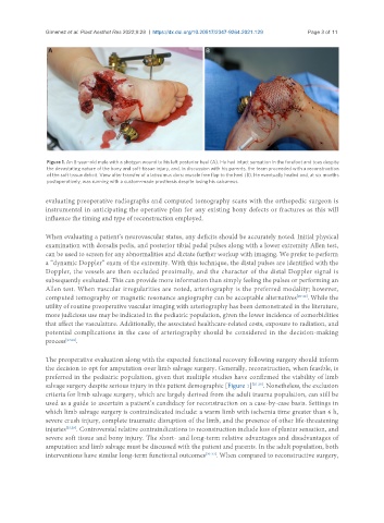
Figure 1. An 8-year-old male with a shotgun wound to his left posterior heel (A). He had intact sensation in the forefoot and toes despite
the devastating nature of the bony and soft-tissue injury, and, in discussion with his parents, the team proceeded with a reconstruction
of the soft tissue deficit. View after transfer of a latissimus dorsi muscle free flap to the heel (B). He eventually healed and, at six months
postoperatively, was running with a custom-made prosthesis despite losing his calcaneus.
evaluating preoperative radiographs and computed tomography scans with the orthopedic surgeon is
instrumental in anticipating the operative plan for any existing bony defects or fractures as this will
influence the timing and type of reconstruction employed.
When evaluating a patient’s neurovascular status, any deficits should be accurately noted. Initial physical
examination with dorsalis pedis, and posterior tibial pedal pulses along with a lower extremity Allen test,
can be used to screen for any abnormalities and dictate further workup with imaging. We prefer to perform
a “dynamic Doppler” exam of the extremity. With this technique, the distal pulses are identified with the
Doppler, the vessels are then occluded proximally, and the character of the distal Doppler signal is
subsequently evaluated. This can provide more information than simply feeling the pulses or performing an
Allen test. When vascular irregularities are noted, arteriography is the preferred modality; however,
computed tomography or magnetic resonance angiography can be acceptable alternatives [20-22] . While the
utility of routine preoperative vascular imaging with arteriography has been demonstrated in the literature,
more judicious use may be indicated in the pediatric population, given the lower incidence of comorbidities
that affect the vasculature. Additionally, the associated healthcare-related costs, exposure to radiation, and
potential complications in the case of arteriography should be considered in the decision-making
process [23,24] .
The preoperative evaluation along with the expected functional recovery following surgery should inform
the decision to opt for amputation over limb salvage surgery. Generally, reconstruction, when feasible, is
preferred in the pediatric population, given that multiple studies have confirmed the viability of limb
salvage surgery despite serious injury in this patient demographic [Figure 1] [25-28] . Nonetheless, the exclusion
criteria for limb salvage surgery, which are largely derived from the adult trauma population, can still be
used as a guide to ascertain a patient’s candidacy for reconstruction on a case-by-case basis. Settings in
which limb salvage surgery is contraindicated include: a warm limb with ischemia time greater than 6 h,
severe crush injury, complete traumatic disruption of the limb, and the presence of other life-threatening
injuries [25,29] . Controversial relative contraindications to reconstruction include loss of plantar sensation, and
severe soft tissue and bony injury. The short- and long-term relative advantages and disadvantages of
amputation and limb salvage must be discussed with the patient and parents. In the adult population, both
interventions have similar long-term functional outcomes [30-33] . When compared to reconstructive surgery,