
Page 48 - Read Online
P. 48
Jabbour et al. Plast Aesthet Res 2021;8:43 https://dx.doi.org/10.20517/2347-9264.2021.59 Page 3 of 12
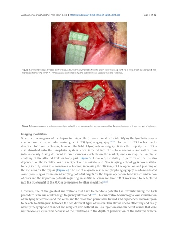
Figure 1. Lymphovenous bypass performed, allowing the lymphatic fluid to drain into the recipient vein. The green background has
markings delineating 1 mm × 1 mm squares demonstrating the submillimeter vessels that are repaired.
Figure 2. Lymphovenous anastomosis performed with a venous coupling device completing the anastomosis without the use of sutures.
Imaging modalities
Since the re-emergence of the bypass technique, the primary modality for identifying the lymphatic vessels
centered on the use of indocyanine green (ICG) lymphangiography [20-22] . The use of ICG has been well-
described for tissue perfusion; however, the field of lymphedema surgery utilizes the property that ICG is
also absorbed into the lymphatic system when injected into the subcutaneous space rather than
intravascularly. Using different infrared cameras available on the market, one can map the lymphatic
anatomy of the affected limb or body part [Figure 3]. However, the ability to perform an LVB is also
dependent on the identification of a recipient vein of suitable size. New imaging technology is now available
to help identify veins in a non-invasive fashion, increasing the efficiency of the operation and planning of
the incisions for the bypass [Figure 4]. The use of magnetic resonance lymphangiography has demonstrated
some promising outcomes in identifying potential targets for the bypass operation; however, consideration
of costs and the impact on patients requiring an additional exam and time off of work need to be factored
into the true benefit of the MR in comparison to other modalities [22,23] .
However, one of the greatest innovations that have tremendous potential in revolutionizing the LVB
procedure is the use of ultra-high frequency ultrasound [24,25] . This innovative technology allows visualization
of the lymphatic vessels and the veins, and the resolution permits the trained and experienced microsurgeon
to be able to distinguish between the two different types of vessels. This allows one to effectively and easily
identify the lymphatic channel and recipient vein without an ICG injection and can detect vessels that were
not previously visualized because of the limitations in the depth of penetration of the infrared camera.