
Page 49 - Read Online
P. 49
Page 4 of 12 Jabbour et al. Plast Aesthet Res 2021;8:43 https://dx.doi.org/10.20517/2347-9264.2021.59
Figure 3. Indocyanine green (ICG) lymphatic mapping demonstrating the linear lymphatic channels that have absorbed the ICG
fluorescent dye. This is a linear pattern or an MD Anderson stage 1 without obvious dermal backflow.
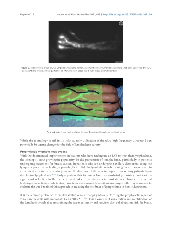
Figure 4. Vein finder device utilized to identify potential targets for recipient veins.
While the technology is still in its infancy, early utilization of the ultra-high frequency ultrasound can
potentially be a game changer for the field of lymphedema surgery.
Prophylactic lymphovenous bypass
With the documented improvements in patients who have undergone an LVB to treat their lymphedema,
the concept is now growing in popularity for the prevention of lymphedema, particularly in patients
undergoing treatment for breast cancer. In patients who are undergoing axillary dissection, using the
lymphatic preventative healing approach (LYMPHA), the lymphatic vessels draining the arm are repaired to
a recipient vein in the axilla to preserve the drainage of the arm in hopes of preventing patients from
developing lymphedema [26-28] . Early reports of this technique have demonstrated promising results with a
significant reduction in the incidence and risks of lymphedema in most studies. However, the actual
technique varies from study to study and from one surgeon to another, and longer follow-up is needed to
evaluate the true benefit of this approach in reducing the incidence of lymphedema in high-risk patients.
It is the authors’ preference to employ axillary reverse mapping when performing the prophylactic repair of
vessels in the axilla with immediate LVB (PREVAIL) . This allows direct visualization and identification of
[29]
the lymphatic vessels that are draining the upper extremity and requires close collaboration with the breast