
Page 51 - Read Online
P. 51
Page 6 of 12 Jabbour et al. Plast Aesthet Res 2021;8:43 https://dx.doi.org/10.20517/2347-9264.2021.59
Figure 5. Axillary reverse mapping using isosulfan blue to identify the lymphatic vessels. The use of the chromophore dye allows
visualization of the lymphatic channels during the axillary dissection, which facilitates the prophylactic repair of vessels in the axilla with
immediate lymphovenous bypass (PREVAIL).
Figure 6. Lymphovenous bypass performed using a double-barrel approach with the anastomosis of two lymphatic vessels into a single
larger caliber recipient vein.
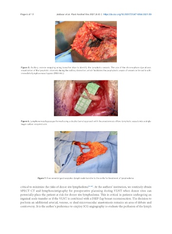
Figure 7. Free omental gastroepiploic lymph node transfer to the axilla for treatment of lymphedema.
critical to minimize the risks of donor site lymphedema [43,44] . At the authors’ institution, we routinely obtain
SPECT-CT and lymphoscintigraphy for preoperative planning during VLNT when donor sites can
potentially place the patient at risk for donor site lymphedema. This is critical in patients undergoing an
inguinal node transfer or if the VLNT is combined with a DIEP flap breast reconstruction. The decision to
perform an additional arterial, venous, or dual microvascular anastomosis remains an area of debate and
controversy. It is the author’s preference to employ ICG angiography to evaluate the perfusion of the lymph