
Page 52 - Read Online
P. 52
Jabbour et al. Plast Aesthet Res 2021;8:43 https://dx.doi.org/10.20517/2347-9264.2021.59 Page 7 of 12
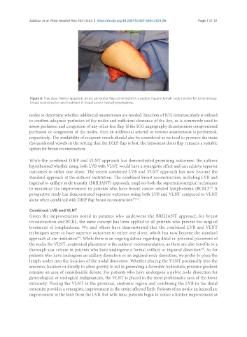
Figure 8. Free deep inferior epigastric artery perforator flap combined with a pedicle inguinal lymph node transfer for simultaneous
breast reconstruction and treatment of breast cancer-related lymphedema.
nodes to determine whether additional anastomoses are needed. Injection of ICG intravascularly is utilized
to confirm adequate perfusion of the nodes and sufficient clearance of the dye, as is commonly used to
assess perfusion and congestion of any other free flap. If the ICG angiography demonstrates compromised
perfusion or congestion of the nodes, then an additional arterial or venous anastomosis is performed,
respectively. The availability of recipient vessels should also be considered as we tend to preserve the main
thoracodorsal vessels in the setting that the DIEP flap is lost; the latissimus dorsi flap remains a suitable
option for breast reconstruction.
While the combined DIEP and VLNT approach has demonstrated promising outcomes, the authors
hypothesized whether using both LVB with VLNT would have a synergistic effect and can achieve superior
outcomes to either one alone. The recent combined LVB and VLNT approach has now become the
standard approach at the authors’ institution. The combined breast reconstruction, including LVB and
inguinal to axillary node transfer (BRILIANT) approach, employs both the supermicrosurgical techniques
[45]
to maximize the improvement in patients who have breast cancer-related lymphedema (BCRL) . A
prospective study has demonstrated superior outcomes using both LVB and VLNT compared to VLNT
alone when combined with DIEP flap breast reconstruction [45,46] .
Combined LVB and VLNT
Given the improvements noted in patients who underwent the BRILIANT approach for breast
reconstruction and BCRL, the same concept has been applied to all patients who present for surgical
treatment of lymphedema. We and others have demonstrated that the combined LVB and VLNT
techniques seem to have superior outcomes to either one alone, which has now become the standard
approach at our institution . While there is an ongoing debate regarding distal vs. proximal placement of
[47]
the nodes for VLNT, anatomical placement is the authors’ recommendation, as there are also benefits to a
[48]
thorough scar release in patients who have undergone a formal axillary or inguinal dissection . So for
patients who have undergone an axillary dissection or an inguinal node dissection, we prefer to place the
lymph nodes into the location of the nodal dissection. Whether placing the VLNT proximally into the
anatomic location or distally to allow gravity to aid in generating a favorable hydrostatic pressure gradient
remains an area of considerable debate. For patients who have undergone a pelvic node dissection for
gynecological or urological malignancies, the VLNT is placed in the most problematic area of the lower
extremity. Placing the VLNT in the proximal, anatomic region and combining the LVB in the distal
extremity provides a synergistic improvement in the entire affected limb. Patients often notice an immediate
improvement in the limb from the LVB, but with time, patients begin to notice a further improvement as