
Page 40 - Read Online
P. 40
Calderwood et al. Plast Aesthet Res 2021;8:40 https://dx.doi.org/10.20517/2347-9264.2021.14 Page 9 of 14
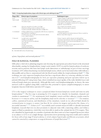
Table 1. Comparing lymphoedema stages with clinical signs and radiological signs [6,11,22]
Stage (ISL) Clinical signs & symptoms Radiological signs
0/Ia • Swelling is not evident, despite impaired lymph transport • Subtle changes in subcutaneous tissue fluid composition
(subclinical) • Asymptomatic or subjective symptoms of limb heaviness • Abnormal lymphatic vessels
• No clinical signs • Fewer lymph nodes than lymphatic vessels
• Interruption of lymphatic vessels with or without distal
lymphatic vessel dilatation
I (mild) • Early accumulation of protein-rich fluid • Less visualized lymphatic vessels
• May have pitting oedema (without fibrosis) • Delay of contrast agent transport
• Oedema subsides with limb elevation within 24 h
II (moderate) • Pitting oedema (may no longer pit as the fibrosis progresses, • Honeycomb pattern
reducing tissue compliance) • Contrast agent accumulation between fat surrounded by
• Oedema does not resolve with limb elevation alone fibrotic tissue (late stage 2)
• Loss of joint flexibility
III (severe) • Lymphostatic elephantiasis • Large, irregular, patchy shaped dermal backflow
• Pitting usually absent • Increased subcutaneous thickness and diffuse fibrosis
• Skin hyperkeratosis/acanthosis • Dilated lymph vessels and damaged lymph vessels even
• Fat deposits with severe fibrosis
• Fibrosis • Tortuous and deep-seated lymph vessels (due to
• Warty overgrowths overgrowth of adipose tissue)
• Marked functional loss
ISL: International Society of Lymphology.
aplasic, hypoplasic and normal patterns [12,19,21] .
ROLE IN SURGICAL PLANNING
MRL plays a vital role in planning surgeries and choosing the appropriate procedure based on the structural
abnormality causing the lymphoedema. Lymph node transfer (LNT) is used for lymphoedema of moderate
severity and in patients who have had lymph node dissections or radiotherapy as part of their oncology
treatment. LNT utilizes functioning vascularized lymph nodes, which can be harvested either with/without a
skin paddle and are then re-anastomosed with the blood vessels within the lymphoedematous limb [37,38] . This
technique not only improves lymphoedema but has a significant effect on reducing cellulitis in limbs.
[38]
Lin et al. used lymphoscintigraphy for both pre-operative planning and post-operative follow-up in
patients following LNT using tissue flaps, which identified increased uptake of the radio-labelled tracer and
reduced lymph stasis post-operatively. Although this study used lymphoscintigraphy, in theory, the same
method could be applied using MRL could have the potential to replace lymphoscintigraphy to assess
lymphatic function both before and after LNT surgery.
LVA is the surgical technique to create a peripheral shunt between lymphatic vessels and veins in early
lymphoedema [5,19] . The first step in planning for LVA surgery is to identify the lymphatic channels and
venules to anastomose [2,28,30] . For anastomosis, both lymphatic vessels and their adjacent venules must be
between 0.3-0.8 mm in calibre, and the lymphatics must not be sclerotic or tortuous [5,10,11,23] . The number,
calibre, anatomical location, and distribution of the lymphatic vessels in the affected limb should be
evaluated prior to surgery to ensure that they fit the criteria for surgery and predict the chance of a
successful outcome [2,3,11,23] . Zeltzer et al. successfully used contrast-enhanced MRL to identify functional
[28]
lymphatic channels that were in close proximity to adjacent veins with a matching calibre, and within a
region of fluid accumulation, which they were then able to mark as the optimal site for LVA surgery. They
also identified fat hypertrophy in a number of patients who then went on to have targeted liposuction either
as an adjunct to LVA or as a single therapy. Liposuction shows promising long-term results and is suitable
for those with at least stage II lymphoedema, in which adipose hypertrophy and fibrosis are present, which
cannot be removed by compression or surgical diversion of lymph fluid alone [39,40] .