
Page 37 - Read Online
P. 37
Page 6 of 14 Calderwood et al. Plast Aesthet Res 2021;8:40 https://dx.doi.org/10.20517/2347-9264.2021.14
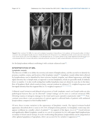
Figure 3. Non-contrast FSE MRL of a man with moderate hyperplasic bilateral lower limb oedema. (a) Increased number of dilated
lymphatic iliac and inguinal trunks (arrows). Bilateral hydrocoele (H) present. (b, c) Dilated lymphatic vessels (arrows) seen at the
lower levels of the limbs and fluid infiltration (I) of subcutaneous fat. (d) Water IDEAL T2 FSE image. Bilateral fluid infiltration (I) of
subcutaneous fat with a honeycomb pattern, moderate epifascial collection (C) and increased thickness of the dermis (arrow) [20] .
for the lymphoedema without combining it with contrast-enhanced scans .
[14]
INTERPRETATION OF MRL
Lymphatic vessels
The first role of MRL is to define the severity and extent of lymphoedema, and the second is to identify the
presence, number, course, and location of the lymphatic vessels . Lymphatic vessels within limbs affected
[2,6]
by lymphoedema can be identified by their tortuous, beaded, irregular, and dilated appearance with high
signal intensity due to lymph stasis, as opposed to normal lymphatics, which are generally small, ill-defined,
fewer in number. It is also worth noting that healthy lymphatics may not always be seen due to the faster
flow of lymphatic fluid, which results in faster contrast washout in enhanced T1-weighted sequences and
less signal intensity than the stagnant fluid in T2-weighted sequences [2-4,7,11,14,15,19,24,26] .
Collateral vessel formation and delayed enhancement of both lymphatic vessels and lymph nodes are also
pathological features that can be observed . Lymph leakage can be seen on contrast-enhanced MRL
[7]
following trauma or iatrogenic damage to the vessel and in post-operative anastomotic leaks [3,4,7,17,29] . Post-
contrast scans show a vast accumulation of contrast agents in affected limbs of patients with unilateral
[15]
lymphoedema compared to their healthy limbs .
Of note, there is some variation in the appearance of lymphatic vessels. The typical tortuous beaded
appearance described above is seen in 80%-90% of lymphoedema patients, but lymphatic vessels may also
appear rectilinear in a smaller cohort of patients [3,18] . In support of this figure, Liu et al. found tortuous
[29]
and significantly dilated lymphatic vessels in 104 out of 123 patients (84.5%) with upper limb lymphoedema
secondary to breast cancer therapy. It is also worth noting that primary lymphoedema can be categorised as
either aplasia/hypoplasia or hyperplasia, which can account for the variation in appearance [13,25] .