
Page 36 - Read Online
P. 36
Calderwood et al. Plast Aesthet Res 2021;8:40 https://dx.doi.org/10.20517/2347-9264.2021.14 Page 5 of 14
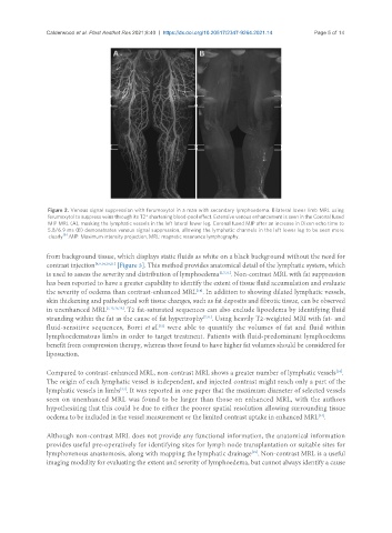
Figure 2. Venous signal suppression with ferumoxytol in a man with secondary lymphoedema. Bilateral lower limb MRL using
ferumoxytol to suppress veins through its T2* shortening blood-pool effect. Extensive venous enhancement is seen in the Coronal fused
MIP MRL (A), masking the lymphatic vessels in the left lateral lower leg. Coronal fused MIP after an increase in Dixon echo time to
5.8/6.9 ms (B) demonstrates venous signal suppression, allowing the lymphatic channels in the left lower leg to be seen more
[6]
clearly . MIP: Maximum intensity projection; MRL: magnetic resonance lymphography.
from background tissue, which displays static fluids as white on a black background without the need for
contrast injection [8,9,14,19,21] [Figure 3]. This method provides anatomical detail of the lymphatic system, which
is used to assess the severity and distribution of lymphoedema [2,7,31] . Non-contrast MRL with fat suppression
has been reported to have a greater capability to identify the extent of tissue fluid accumulation and evaluate
[14]
the severity of oedema than contrast-enhanced MRL . In addition to showing dilated lymphatic vessels,
skin thickening and pathological soft tissue changes, such as fat deposits and fibrotic tissue, can be observed
in unenhanced MRL [7,13,25,28] . T2 fat-saturated sequences can also exclude lipoedema by identifying fluid
stranding within the fat as the cause of fat hypertrophy [7,31] . Using heavily T2-weighted MRI with fat- and
fluid-sensitive sequences, Borri et al. were able to quantify the volumes of fat and fluid within
[32]
lymphoedematous limbs in order to target treatment. Patients with fluid-predominant lymphoedema
benefit from compression therapy, whereas those found to have higher fat volumes should be considered for
liposuction.
Compared to contrast-enhanced MRL, non-contrast MRL shows a greater number of lymphatic vessels .
[14]
The origin of each lymphatic vessel is independent, and injected contrast might reach only a part of the
lymphatic vessels in limbs . It was reported in one paper that the maximum diameter of selected vessels
[33]
seen on unenhanced MRL was found to be larger than those on enhanced MRL, with the authors
hypothesizing that this could be due to either the poorer spatial resolution allowing surrounding tissue
oedema to be included in the vessel measurement or the limited contrast uptake in enhanced MRL .
[14]
Although non-contrast MRL does not provide any functional information, the anatomical information
provides useful pre-operatively for identifying sites for lymph node transplantation or suitable sites for
lymphovenous anastomosis, along with mapping the lymphatic drainage . Non-contrast MRL is a useful
[19]
imaging modality for evaluating the extent and severity of lymphoedema, but cannot always identify a cause