
Page 35 - Read Online
P. 35
Page 4 of 14 Calderwood et al. Plast Aesthet Res 2021;8:40 https://dx.doi.org/10.20517/2347-9264.2021.14
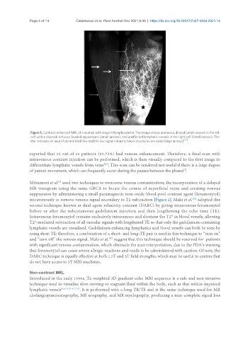
Figure 1. Contrast-enhanced MRL of a woman with stage III lymphoedema. The image shows numerous dilated lymph vessels in the left
calf with a classical tortuous beaded appearance (small arrows), and unaffected lymphatic vessels in the right calf (small arrows). The
star indicates an area of dermal backflow and the low signal intensity linear structures are veins (large arrows) [24] .
reported that 15 out of 16 patients (93.75%) had venous enhancement. Therefore, a final scan with
intravenous contrast injection can be performed, which is then visually compared to the first image to
[2,3]
differentiate lymphatic vessels from veins . This scan can be rendered not useful if there is a large degree
[2]
of patient movement, which can frequently occur during the pauses between the phases .
Mitsumori et al. used two techniques to overcome venous contamination; the incorporation of a delayed
[6]
MR venogram using the same GBCA to locate the course of superficial veins and creating venous
suppression by administering a small paramagnetic iron-oxide blood-pool contrast agent (ferumoxytol)
[30]
intravenously to remove venous signal secondary to T2 subtraction [Figure 2]. Maki et al. adopted this
second technique known as dual-agent relaxivity contrast (DARC) by giving intravenous ferumoxytol
before or after the subcutaneous gadolinium injection and then lengthening the echo time (TE).
Intravenous ferumoxytol remains exclusively intravenous and shortens the T2* in blood vessels, allowing
T2*-mediated subtraction of all vascular signals with lengthened TE so that only the gadolinium-containing
lymphatic vessels are visualised. Gadolinium-enhancing lymphatics and blood vessels can both be seen by
using short TE; therefore, a combination of a short- and long-TE pair is used in this technique to “turn on”
[30]
and “turn off” the venous signal. Maki et al. suggest that this technique should be reserved for patients
with significant venous contamination, which obstructs the scan interpretation, due to the FDA’s warning
that ferumoxytol can cause severe allergic reactions and needs to be administered with caution. Of note, the
DARC technique is equally effective at both 1.5T and 3T field strengths, which may be useful to centres that
do not have access to 3T MRI machines.
Non-contrast MRL
Introduced in the early 1990s, T2-weighted 3D gradient-echo MRI sequence is a safe and non-invasive
technique used to visualise slow-moving or stagnant fluid within the body, such as that within impaired
lymphatic vessels [8,9,14,15,19,21,31] . It is performed with a long TR/TE and is the same technique used for MR
cholangiopancreatography, MR urography, and MR myelography, producing a near-complete signal loss