
Page 12 - Read Online
P. 12
Page 6 of 11 Drobot et al. Plast Aesthet Res 2021;8:30 https://dx.doi.org/10.20517/2347-9264.2021.19
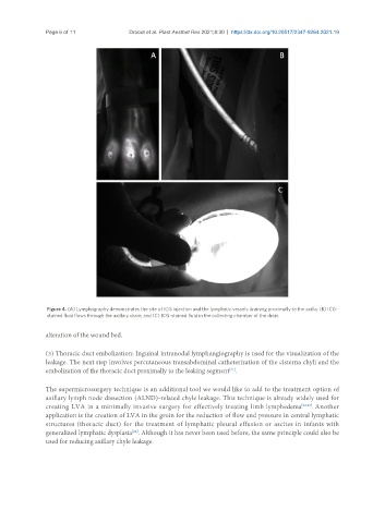
Figure 4. (A) Lymphography demonstrates the site of ICG injection and the lymphatic vessels draining proximally to the axilla; (B) ICG-
stained fluid flows through the axillary drain; and (C) ICG-stained fluid in the collecting chamber of the drain.
alteration of the wound bed.
(3) Thoracic duct embolization: Inguinal intranodal lymphangiography is used for the visualization of the
leakage. The next step involves percutaneous transabdominal catheterization of the cisterna chyli and the
embolization of the thoracic duct proximally to the leaking segment .
[21]
The supermicrosurgery technique is an additional tool we would like to add to the treatment option of
axillary lymph node dissection (ALND)-related chyle leakage. This technique is already widely used for
creating LVA in a minimally invasive surgery for effectively treating limb lymphedema [22,23] . Another
application is the creation of LVA in the groin for the reduction of flow and pressure in central lymphatic
structures (thoracic duct) for the treatment of lymphatic pleural effusion or ascites in infants with
generalized lymphatic dysplasia . Although it has never been used before, the same principle could also be
[24]
used for reducing axillary chyle leakage.