
Page 9 - Read Online
P. 9
Drobot et al. Plast Aesthet Res 2021;8:30 https://dx.doi.org/10.20517/2347-9264.2021.19 Page 3 of 11
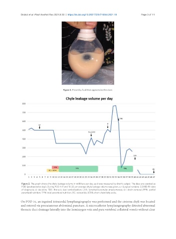
Figure 1. The milky fluid that appeared in the drain.
Figure 2. The graph shows the chyle leakage volume in milliliters per day, as it was measured by drain’s output. The days are counted as
POD (postoperative day). During POD 4-9 and 10-20, an average chyle leakage volume was given. s.r: Surgical revision; COVID-19: date
of diagnosis; d: decision; TDE: thoracic duct embolization; LVA: lymphaticovenular anastomoses; d.r: drain removal; PPN: partial
parenteral nutrition; TPN: total parenteral nutrition; OC: octreotide; SCFA: short chain fatty acids.
On POD 34, an inguinal intranodal lymphangiography was performed and the cisterna chyli was located
and entered via percutaneous abdominal puncture. A microcatheter lymphangiography detected abnormal
thoracic duct drainage laterally into the hemiazygos vein and para vertebral collateral vessels without clear