
Page 11 - Read Online
P. 11
Drobot et al. Plast Aesthet Res 2021;8:30 https://dx.doi.org/10.20517/2347-9264.2021.19 Page 5 of 11
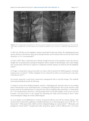
Figure 3. (A) A lymphographic demonstration of the thoracic duct, its draining branch (B.H) into the hemiazygos vein (H.V), and the
small collateral lymph vessel (S.C) that originates from its distal end; and (B) a coil (C) used for the embolization of the distal thoracic
duct.
(3) Bed rest: The flow in the lymphatic system is generated by physical activity. By minimizing physical
activity, the flow in the thoracic duct and its leaking branches can be reduced. Bed rest with the head of bed
elevated to 30°-45° is recommended .
[9]
(4) Diet: A MCT diet is commonly used, with the thought that most of its absorption crosses the mucosa,
straight into the portal system, sparing the lymphatic system. Despite its disadvantages, when chyle leakage
does not decreases, TPN may be required to completely avoid the entrance of nutrients into the lymphatic
system .
[10]
(5) Drugs: A somatostatin analog (octerotide) can reduce chyle production by inhibiting gastric, intestinal,
and pancreatic secretions . Further, lymphatic flow is reduced by its effect on smooth muscle contraction
[11]
in the thoracic duct .
[12]
An invasive approach is used when conservative management fails to stop the leakage. The available
interventional methods include the following:
(1) Surgical reexploration: Finding lymphatic vessels is a challenging task, especially when the surrounding
tissue is inflamed due to the extravasated chyle. Localizing the leaking thoracic duct and its branches could
become easier by the administration of a lipophilic dye such as methylene blue, patent blue, or Sudan black
B [13,14] . For ameliorating its detection, a fasting patient with a lucid chyle leakage could receive a fat-rich
formula a few hours prior to the surgery for whitening the chyle and increasing its flow . After
[15]
demonstrating the leakage, the vessels could be ligated.
(2) Local agents: Local inflammatory reaction can generate fibrosis and seal the chylous leakage. This
reaction may be induced by applying an intralesional injection of sclerosing agents such as OK-432 or
[16]
tetracycline . Other agents that could be used for sealing the leakage are cyanoacrylate [18,19] , fibrin glue, and
[17]
polyglactin (Vicryl) mesh . Reexploration after using those agents could be difficult due to the tissue
[20]