
Page 13 - Read Online
P. 13
Page 6 of 24 Reilly et al. Plast Aesthet Res 2021;8:2 I http://dx.doi.org/10.20517/2347-9264.2020.153
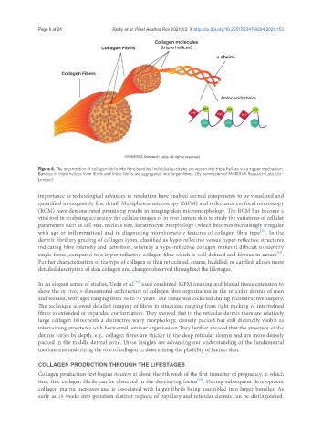
Figure 4. The organization of collagen fibrils into fibre bundles. Individual α-chains are woven into triple helices via a zipper mechanism.
Bundles of triple helices form fibrils and these fibrils are aggregated into larger fibres. (By permission of MINERVA Research Labs Ltd -
London)
importance as technological advances in resolution have enabled dermal components to be visualized and
quantified in exquisitely fine detail. Multiphoton microscopy (MPM) and reflectance confocal microscopy
(RCM) have demonstrated promising results in imaging skin micromorphology. The RCM has become a
vital tool in analysing accurately the cellular images of in vivo human skin to study the variations of cellular
parameters such as cell size, nucleus size, keratinocyte morphology (which becomes increasingly irregular
[31]
with age or inflammation) and in diagnosing morphometric features of collagen fibre type . In the
dermis fibrillary grading of collagen types, classified as hypo-reflective versus hyper-reflective structures
indicating fibre intensity and definition, whereby a hypo-reflective collagen makes it difficult to identify
[32]
single fibres, compared to a hyper-reflective collagen fibre which is well defined and fibrous in nature .
Further characterisation of the type of collagen as thin reticulated, coarse, huddled, or curdled, allows more
detailed description of skin collagen and changes observed throughout the lifestages.
[33]
In an elegant series of studies, Ueda et al. used combined MPM imaging and biaxial tissue extension to
show the in vivo, 3-dimensional architecture of collagen fibre organization in the reticular dermis of men
and women, with ages ranging from 36 to 75 years. The tissue was collected during reconstructive surgery.
The technique allowed detailed imaging of fibres in situations ranging from tight packing of intertwined
fibres to extended or expanded conformation. They showed that in the reticular dermis there are relatively
large collagen fibres with a distinctive wavy morphology, densely packed but still distinctly visible as
intertwining structures with horizontal laminar organization They further showed that the structure of the
dermis varies by depth, e.g., collagen fibres are thicker in the deep reticular dermis and are more densely
packed in the middle dermal zone. These insights are advancing our understanding of the fundamental
mechanisms underlying the role of collagen in determining the pliability of human skin.
COLLAGEN PRODUCTION THROUGH THE LIFESTAGES
Collagen production first begins in utero at about the 5th week of the first trimester of pregnancy, at which
[34]
time fine collagen fibrils can be observed in the developing foetus . During subsequent development
collagen matrix increases and is associated with larger fibrils being assembled into larger bundles. As
early as 15 weeks into gestation distinct regions of papillary and reticular dermis can be distinguished.