
Page 11 - Read Online
P. 11
Page 4 of 24 Reilly et al. Plast Aesthet Res 2021;8:2 I http://dx.doi.org/10.20517/2347-9264.2020.153
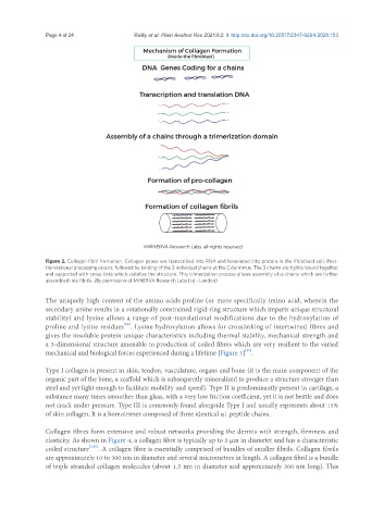
Figure 2. Collagen fibril formation. Collagen genes are transcribed into RNA and translated into protein in the fibroblast cell. Post-
translational processing occurs, followed by binding of the 3 individual chains at the C-terminus. The 3 chains are tightly bound together
and supported with cross-links which stabilise the structure. This trimerization process allows assembly of α-chains which are further
assembled into fibrils. (By permission of MINERVA Research Labs Ltd - London)
The uniquely high content of the amino acids proline (or more specifically imino acid, wherein the
secondary amine results in a rotationally constrained rigid-ring structure which imparts unique structural
stability) and lysine allows a range of post-translational modifications due to the hydroxylation of
[24]
proline and lysine residues . Lysine hydroxylation allows for crosslinking of intertwined fibres and
gives the insoluble protein unique characteristics including thermal stability, mechanical strength and
a 3-dimensional structure amenable to production of coiled fibres which are very resilient to the varied
[25]
mechanical and biological forces experienced during a lifetime [Figure 3] .
Type I collagen is present in skin, tendon, vasculature, organs and bone (it is the main component of the
organic part of the bone, a scaffold which is subsequently mineralized to produce a structure stronger than
steel and yet light enough to facilitate mobility and speed). Type II is predominantly present in cartilage, a
substance many times smoother than glass, with a very low friction coefficient, yet it is not brittle and does
not crack under pressure. Type III is commonly found alongside Type I and usually represents about 15%
of skin collagen. It is a homotrimer composed of three identical α1 peptide chains.
Collagen fibres form extensive and robust networks providing the dermis with strength, firmness and
elasticity. As shown in Figure 4, a collagen fibre is typically up to 3 μm in diameter and has a characteristic
coiled structure [2,26] . A collagen fibre is essentially comprised of bundles of smaller fibrils. Collagen fibrils
are approximately 10 to 300 nm in diameter and several micrometres in length. A collagen fibril is a bundle
of triple stranded collagen molecules (about 1.5 nm in diameter and approximately 300 nm long). This