
Page 41 - Read Online
P. 41
Hara et al. Plast Aesthet Res 2023;10:42 https://dx.doi.org/10.20517/2347-9264.2023.11 Page 3 of 11
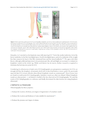
Figure 1. Multi-point indocyanine green (ICG) lymphography (originally reported as multi-lymphosome lymphography). (A) Schematic
illustration of multi-point ICG lymphography. The crosses indicate the injection points of ICG (the first web space, the proximal point of
the lateral malleolus, and the lateral midline point at the level of the superior border of the patella). The red line indicates the lymphatic
vessel in the Saphenous lymphatic area, the green line indicates the lymphatic vessel in the Lateral calf lymphatic area, and the blue line
indicates the lymphatic vessel in the Lateral thigh lymphatic area. When performing lymphatic ultrasound, we apply the probe so that
the lymphatic vessel and the probe are at right angles, imagining the running of these lymphatic vessels (-●); (B) Clinical pictures of
multi-point ICG lymphography.
Shinaoka et al. reported on the lymphatic map with injecting ICG “below the medial malleolus, below the
lateral malleolus, in the first interdigital space, fourth interdigital space, and at the midpoint of the straight
line that connects the head of the fifth metatarsal bone and the lateral malleolus” . We agree with their
[42]
theory, although additional injections in the proximal part (the calf and thigh) are necessary to evaluate the
lymphatic vessels in the whole leg. While each theory has its merits, further research is required to
determine the best injection point for ICG.
Considering the effectiveness of multi-point ICG lymphography as a preoperative examination for LVA, we
strongly feel that the lymphatic ultrasound, which will be described below, is more useful. We previously
reported that LVA is most effective when dilated lymphatic vessels are anastomosed . Even if some lines
[3]
are found on multi-point ICG lymphography, lymphatic vessels are often not dilated. Dilated lymphatic
vessels are often present 1-2 cm lateral or medial to the line observed on ICG lymphography. We used the
results of ICG lymphography as a reference and finally determined the skin incision site using lymphatic
ultrasound.
LYMPHATIC ULTRASOUND
Ultrasonography has three purposes.
1. Evaluate the location, thickness, and degree of degeneration of lymphatic vessels.
2. Evaluate the location and thickness of veins suitable for anastomosis .
[43]
3. Evaluate the presence and degree of edema.