
Page 45 - Read Online
P. 45
Hara et al. Plast Aesthet Res 2023;10:42 https://dx.doi.org/10.20517/2347-9264.2023.11 Page 7 of 11
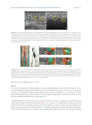
Figure 5. Example of lymphatic ultrasound at the lymphedematous medial calf using different kinds of probes. Blue circles indicate veins
and yellow circles indicate lymphatic vessels. White arrowhead indicates a depth of 5 mm. (A) Ultrasonogram with an 18 MHz linear
probe (Noblus EUP-L65; Hitachi Medical Corp., Tokyo, Japan). One vein and one lymphatic vessel can be observed. The dashed circle
indicates the lymphatic vessel, which we found only after observing it with a 33MHz probe; (B) Ultrasonogram with a 33 MHz linear
probe (Aplio i900, Canon Medical Systems Corp., Tokyo, Japan). One vein and three lymphatic vessels are recognized. There are two
lymphatic vessels in the central yellow circle. Compared to A, it can be seen that ultrasound is attenuated in areas deeper than 5 mm,
making it difficult to observe.
Figure 6. Case 1. A 52-year-old woman with secondary lymphedema in the legs. Black lines and numbers indicate skin incisions in
lymphaticovenous anastomosis (LVA). (A) Clinical pictures and results of multi-point indocyanine green (ICG) lymphography. Cross
marks indicate ICG injection sites; (B) Image of lymphoscintigraphy. Dermal backflow is observed in the left thigh. Additionally, slight
dermal backflow is observed around the right inguinal lymph node; (C) Ultrasound findings and corresponding intraoperative findings
during LVA. Blue circles indicate veins and yellow circles indicate lymph vessels. At each site, lymphatic vessels and veins consistent
with ultrasonographic findings are observed. V: vein; L: lymphatic vessel.
perform LVA at a single site was 25-35 min.
Case 2
A 92-year-old woman. She had had primary lower extremity lymphedema since birth. At the age of 90, she
developed idiopathic chylous pleural effusion, which resolved spontaneously. At the age of 91, she had a
recurrence of idiopathic chylous pleural effusion and visited our hospital for the treatment of lower
extremity lymphedema and chylous pleural effusion. Lymphoscintigraphy showed isotope retention in the
left lower extremity and left thoracic cavity [Figure 7] .
[52]
Considering that the isotope injected into the feet leaked into the thoracic cavity, it was possible that leg
compression therapy would increase pleural effusion. As a result of the multidisciplinary discussion about
treatment methods, we decided to perform LVA in the leg first, create an escape route for the lymph, and
then perform leg compression therapy [Figure 7]. Lymphatic ultrasound revealed many dilated lymphatic
vessels. When a skin incision was made on the proximal thigh, cloudy chyle flowed out vigorously, making