
Page 10 - Read Online
P. 10
Men et al. Reconstruction of keloid defect with the rectus abdominis myocutaneous flap
A B
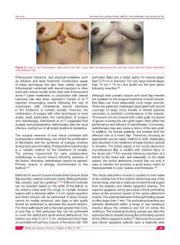
Figure 6: Case 2. (A) Preoperative chest keloid and (B) 1 year after the keloid resection and the repair with the rectus abdominis
myocutaneous flap
5-flourouracil injections, and physical modalities such perforator flaps are a better option for keloids larger
as radiation and laser treatment. Combination usage than 5-10 cm in diameter. For very large keloids larger
of these techniques has also been widely reported. than 10 cm × 10 cm, skin grafts are the best option
5-flourouracil combined with steroid injections is often following resection. [5]
used with clinical results better than with 5-flourouracil
alone. Laser treatments in conjunction with steroid Although both primary closure and local flap transfer
[8]
injections has also been reported. Careta et al. [10] are suitable for the surgical treatment of small keloids,
[9]
reported encouraging results following the use of free flaps can more adequately cover larger wounds.
cryosurgery with intralesional steroid injections There are particular challenges associated with wound
in the treatment of earlobe keloids. However, the coverage of large chest keloids in female patients
combination of surgery with other techniques is more secondary to aesthetic considerations of the breasts.
widely used, particularly the combination of surgery The wound can be covered with a skin graft, but layers
and radiotherapy. Mankowski et al. [11] suggested that of gauze covering the skin graft region often affect the
surgery and postoperative radiotherapy was the most performance and efficacy of radiotherapy. Conversely,
effective method out of all keloid treatment modalities. radiotherapy may also cause a failure of the skin graft.
In addition, for female patients, the breasts limit the
The surgical resection of scar tissue combined with effective use of a chest flap. Therefore, choosing an
postoperative radiotherapy can inhibit the proliferation appropriate wound repair method is both challenging
of fibroblasts and the synthesis of collagen proteins and important in the treatment of large thoracic keloids
during early wound healing. Postoperative radiotherapy in females. The blood supply to the rectus abdominis
is a reliable method for the treatment of keloids. myocutaneous flap is reliable with minimal injury to
The primary requirement for early postoperative the donor site. [17] For wounds following resection of a
radiotherapy is wound closure following resection of keloid on the chest wall, and especially on the distal
the keloid; otherwise, radiotherapy cannot be applied. aspect, the rectus abdominis muscle flap not only is
Primary closure is strongly recommended in this easy to transfer but provides a good tissue match to
method. [12] therecipientsite in color, texture and thickness. [18]
Methods for wound closure include direct closure, local The rectus abdominis muscle is located on both sides
flap transfer, internal mammary artery (IMA) perforator of the median line of the anterior abdominal wall of the
flap transfer, and skin grafting. [13-16] Different methods human body, and has a relatively constant blood supply
can be selected based on the width of the keloid on from the superior and inferior epigastric arteries. The
the anterior chest wall. For single or multiple isolated superior epigastric artery generates a thick perforating
lesions with a diameter within 1-3 cm, keloid resection artery at the proximal aspect of the rectus abdominis
and direct closure can be performed. If direct closure muscle. The external diameter of this perforating vessel
cannot be readily achieved, skin flaps or skin grafts is often larger than 1 mm. The perforating branches are
should be performed to decrease the wound tension. primarily distributed within a range of one tendinous
For chest wall lesions with a diameter greater than 3 cm, insetion above the umbilicus and 8.0 cm below the
a local flap or IMA perforator flap can be performed umbilicus. The medial perforating vessels are often the
to cover the defect and avoid wound dehiscence. For dominant blood vessels among the perforating vessels
lesions less than 5 cm × 5 cm, randomized local flaps of the inferior epigastric artery. [19] Because the superior
are possible with primary closure of the donor site. IMA and inferior epigastric arteries have a relatively wide
Plastic and Aesthetic Research ¦ Volume 4 ¦ May 26, 2017 89