
Page 8 - Read Online
P. 8
Men et al. Reconstruction of keloid defect with the rectus abdominis myocutaneous flap
females in whom the lesions frequently involve the
[2]
breast and other peripheral organs. Many treatment
methods have been reported including triple therapy in
which surgery is combined with steroid injections and
silicone sheet application. While surgery combined
[3]
with radiotherapy is effective in the treatment of
keloids, the wound cannot be directly closed following
[4]
resection of keloids greater than 10 cm × 10 cm in
width and length on the chest wall. In women, given
the presence of breast tissue, it can be even more
difficult to employ primary closure. Skin grafting would
delay radiotherapy. Therefore, for large defects
[5]
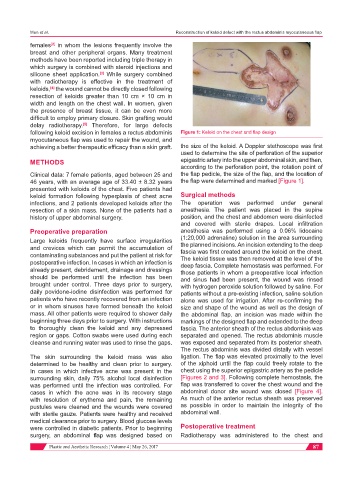
following keloid excision in females a rectus abdominis Figure 1: Keloid on the chest and flap design
myocutaneous flap was used to repair the wound, and
achieving a better therapeutic efficacy than a skin graft. the size of the keloid. A Doppler stethoscope was first
used to determine the site of perforation of the superior
METHODS epigastric artery into the upper abdominal skin, and then,
according to the perforation point, the rotation point of
Clinical data: 7 female patients, aged between 25 and the flap pedicle, the size of the flap, and the location of
46 years, with an average age of 33.40 ± 8.32 years the flap were determined and marked [Figure 1].
presented with keloids of the chest. Five patients had
keloid formation following hyperplasia of chest acne Surgical methods
infections, and 2 patients developed keloids after the The operation was performed under general
resection of a skin mass. None of the patients had a anesthesia. The patient was placed in the supine
history of upper abdominal surgery. position, and the chest and abdomen were disinfected
and covered with sterile drapes. Local infiltration
Preoperative preparation anesthesia was performed using a 0.06% lidocaine
Large keloids frequently have surface irregularities (1:20,000 adrenaline) solution in the area surrounding
the planned incisions. An incision extending to the deep
and crevices which can permit the accumulation of fascia was first created around the keloid on the chest.
contaminating substances and put the patient at risk for The keloid tissue was then removed at the level of the
postoperative infection. In cases in which an infection is deep fascia. Complete hemostasis was performed. For
already present, debridement, drainage and dressings those patients in whom a preoperative local infection
should be performed until the infection has been and sinus had been present, the wound was rinsed
brought under control. Three days prior to surgery, with hydrogen peroxide solution followed by saline. For
daily povidone-iodine disinfection was performed for patients without a pre-existing infection, saline solution
patients who have recently recovered from an infection alone was used for irrigation. After re-confirming the
or in whom sinuses have formed beneath the keloid size and shape of the wound as well as the design of
mass. All other patients were required to shower daily the abdominal flap, an incision was made within the
beginning three days prior to surgery. With instructions markings of the designed flap and extended to the deep
to thoroughly clean the keloid and any depressed fascia. The anterior sheath of the rectus abdominis was
region or gaps. Cotton swabs were used during each separated and opened. The rectus abdominis muscle
cleanse and running water was used to rinse the gaps. was exposed and separated from its posterior sheath.
The rectus abdominis was divided distally with vessel
The skin surrounding the keloid mass was also ligation. The flap was elevated proximally to the level
determined to be healthy and clean prior to surgery. of the xiphoid until the flap could freely rotate to the
In cases in which infective acne was present in the chest using the superior epigastric artery as the pedicle
surrounding skin, daily 75% alcohol local disinfection [Figures 2 and 3]. Following complete hemostasis, the
was performed until the infection was controlled. For flap was transferred to cover the chest wound and the
cases in which the acne was in its recovery stage abdominal donor site wound was closed [Figure 4].
with resolution of erythema and pain, the remaining As much of the anterior rectus sheath was preserved
pustules were cleaned and the wounds were covered as possible in order to maintain the integrity of the
with sterile gauze. Patients were healthy and received abdominal wall.
medical clearance prior to surgery. Blood glucose levels
were controlled in diabetic patients. Prior to beginning Postoperative treatment
surgery, an abdominal flap was designed based on Radiotherapy was administered to the chest and
Plastic and Aesthetic Research ¦ Volume 4 ¦ May 26, 2017 87