
Page 9 - Read Online
P. 9
Men et al. Reconstruction of keloid defect with the rectus abdominis myocutaneous flap
Figure 2: After the formation of the myocutaneous flap of the rectus Figure 3: The pedicle with the perforating vessels of the superior
abdominis epigastric artery
with early radiotherapy to treat 7 cases of large
keloids on the female chest. All flaps survived, and the
incisions healed during the primary stage without any
cases of infection, dehiscence or other complications.
All patients completed their courses of thoracic and
abdominal radiotherapy. Follow-up was conducted
for 10-14 months (average of 12 months) and
demonstrated that all incisions healed well without any
cases of keloid recurrence. Furthermore, the patients
were satisfied with the shape of the chest and abdomen
[Figures 5 and 6].
Figure 4: Closed chest wound 2 weeks after flap transfer DISCUSSION
abdominal donor site on the first and seventh Keloids occur in the skin and can expand rapidly
postoperative days; each dose was 900 cGy for a towards surrounding normal tissue. Keloids are
total dose of 1,800 cGy. The wound was checked and pathologically composed of collagen fibers and
cleaned every three days following surgery and sutures often protrude clinically from the skin. They often
were removed 14 days postoperatively. A compression occur epidemiologically in young people, especially
garment was used 1 month after suture removing until in females. [6,7] Keloids frequently form on the chest,
the wound scar become pale and flat. shoulder, and lower mandible of the face secondary
to acne.
RESULTS
Various therapeutic management techniques have been
From January 2015 to March 2016, the rectus reported in literature including conventional surgery,
abdominis myocutaneous flap was used combined cryosurgery, medical therapy including steroid and
A B
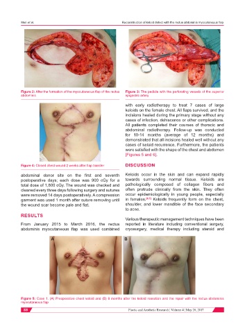
Figure 5: Case 1. (A) Preoperative chest keloid and (B) 8 months after the keloid resection and the repair with the rectus abdominis
myocutaneous flap
88 Plastic and Aesthetic Research ¦ Volume 4 ¦ May 26, 2017