
Page 60 - Read Online
P. 60
Page 8 of 17 Cevallos et al. Plast Aesthet Res 2023;10:30 https://dx.doi.org/10.20517/2347-9264.2023.01
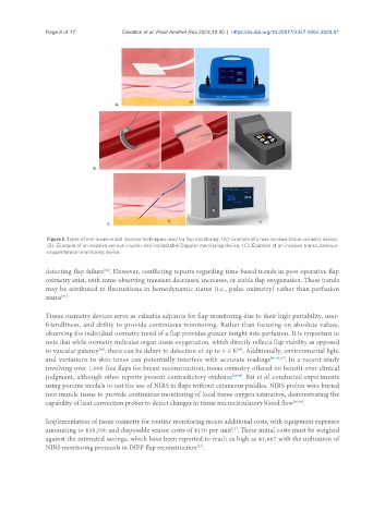
Figure 1. Types of non-invasive and invasive techniques used for flap monitoring. (A): Example of a non-invasive tissue oximetry device;
(B): Example of an invasive venous coupler and implantable Doppler monitoring device; (C): Example of an invasive transcutaneous
oxygen tension monitoring device.
detecting flap failure . However, conflicting reports regarding time-based trends in post-operative flap
[84]
oximetry exist, with some observing transient decreases, increases, or stable flap oxygenation. These trends
may be attributed to fluctuations in hemodynamic status (i.e., pulse oximetry) rather than perfusion
[85]
status .
Tissue oximetry devices serve as valuable adjuncts for flap monitoring due to their high portability, user-
friendliness, and ability to provide continuous monitoring. Rather than focusing on absolute values,
observing the individual oximetry trend of a flap provides greater insight into perfusion. It is important to
note that while oximetry indicates organ tissue oxygenation, which directly reflects flap viability as opposed
to vascular patency , there can be delays in detection of up to 1-5 h . Additionally, environmental light
[80]
[86]
and variations in skin tones can potentially interfere with accurate readings [40-41,87] . In a recent study
involving over 1,000 free flaps for breast reconstruction, tissue oximetry offered no benefit over clinical
judgment, although other reports present contradictory evidence [88,89] . Bai et al. conducted experiments
using porcine models to test the use of NIRS in flaps without cutaneous paddles. NIRS probes were buried
into muscle tissue to provide continuous monitoring of local tissue oxygen saturation, demonstrating the
capability of heat convection probes to detect changes in tissue microcirculatory blood flow [43,90] .
Implementation of tissue oximetry for routine monitoring incurs additional costs, with equipment expenses
[13]
amounting to $16,500 and disposable sensor costs of $150 per unit . These initial costs must be weighed
against the estimated savings, which have been reported to reach as high as $1,667 with the utilization of
NIRS monitoring protocols in DIEP flap reconstruction .
[91]