
Page 58 - Read Online
P. 58
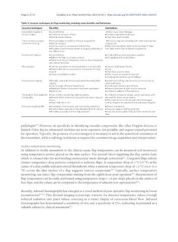
Page 6 of 17            Cevallos et al. Plast Aesthet Res 2023;10:30  https://dx.doi.org/10.20517/2347-9264.2023.01

               Table 3. Invasive techniques for flap monitoring, including main benefits and limitations
                Invasive technique  Benefits                           Limitations
                Implantable doppler &   ● Cost effective                   ● May cause vessel damage
                venous coupler    ● Usable on all types of flaps       ● Complex operative technique
                                  ● Ease in signal reading             ● High false positive rates
                Transcutaneous oxygen   ● Very sensitive to declines in tissue oxygenation       ● Invasive, requires tunneling with some extra dermal
                tension monitoring  ● Easy to monitor                  components
                                  ● Can be used for monitoring of buried flaps   ● May be unreliable in large cutaneous flaps or flaps
                                  ● Provides a direct measurement of oxygen availability at  with large within-flap temperature gradients
                                  the cellular level
                Biochemical markers  ● Cost effective                  ● Limited efficacy among diabetic patients
                                  ● Reflect the flap’s local environment   ● Not applicable for buried flaps
                                  ● Patterns of known metabolites exist to direct thresholds
                                  and clinical decisions
                Microdialysis     ● Can be used when clinical examination is not possible   ● Causes local tissue trauma
                                  ● Detects flap failure hours before it becomes clinically   ● Cost
                                  evident                              ● High false-positive rates
                                  ● Gives a quantitative metric        ● May require an equilibrium period
                                                                       ● Securing the catheter can be difficult
                Fluorescence imaging  ● Broadly used with clinical applications beyond plastic   ● Additional staffing requirements are not as high as
                                  surgery                              other techniques
                                  ● Can leverage different fluorophores    ● Continuous monitoring is difficult
                                  ● Handheld devices and probes have been developed   ● Repeat injections of dyes may be required
                                  ● Ease of use                        ● Limited circulation of fluorophores
                Technetium-99m sestamibi  ● Can monitor muscle flap viability reliably    ● Limited to a majority of case reports and series with
                scintigraphy      ● Reveals distal ischemia or hypoperfused area in muscle  the need for more robust studies
                                  tissue                               ● May need to be combined with other techniques,
                                  ● Short half-life of ~6 h            such as Doppler, to visualize the blood vessel integrity
                Perfusion-weighted MRI  ● Is relatively non-invasive, and risks to the patient are   ● Resource intensive
                                  limited to those imposed by the administration of contrast  ● Requires support staff at each step
                                  ● Provides a picture of entire-flap perfusion  ● Requires serial studies if flap is to be monitored over
                                                                       time


               pathologies . However, its specificity in identifying vascular compromise, like other Doppler devices, is
                         [54]
               limited. Color duplex ultrasound machines are more expensive, less portable, and require trained personnel
               for operation. Typically, the presence of a microsurgeon is necessary to aid in the anatomical orientation of
               the transmitter, while a radiology technician is required for consistent image acquisition and interpretation.

               Surface temperature monitoring
               In addition to tactile assessment in the clinical exam, flap temperature can be measured and monitored
               using temperature probes placed on the skin surface. The arterial blood supplying the flap carries heat,
                                                                                   [63]
               which is released into the surrounding extravascular tissue through convection . Congested flaps exhibit
               distinct temperature drop patterns compared to ischemic flaps. A temperature drop of 3 °C (37 °F) at the
               center of a skin paddle indicates arterial thrombosis, while a uniform temperature drop of 1-2 °C (33.8-35.6
               °F) across the skin surface of a flap suggests venous compromise . Typically, surface temperature
                                                                           [64]
                                                                                         [65]
               monitoring can detect flap compromise starting from the eighth hour post-operation . Measurement of
               flap temperature can be easily performed using inexpensive strips (~1$ per strip) placed on the surface of
                                                                                            [66]
               free flaps, and the values can be compared to the temperature of adjacent non-operated skin .

               Recently, infrared thermography has emerged as a novel method of post-operative flap monitoring in breast
               reconstruction [67,68] . This thermal imaging technology converts the detected temperature values through
               infrared radiation into pixel values, resulting in a visual display of cutaneous blood flow. Infrared
               thermography has demonstrated a sensitivity of 96% and a specificity of 75%, indicating its potential as a
               valuable adjunct to clinical assessment .
                                               [67]